


 Foundational characteristics of cancer include proliferation, angiogenesis, migration, evasion of apoptosis, and cellular immortality. Find key markers for these cellular processes and antibodies to detect them.
 The SUMOplot™ Analysis Program predicts and scores sumoylation sites in your protein. SUMOylation is a post-translational modification involved in various cellular processes, such as nuclear-cytosolic transport, transcriptional regulation, apoptosis, protein stability, response to stress, and progression through the cell cycle.
 The Autophagy Receptor Motif Plotter predicts and scores autophagy receptor binding sites in your protein. Identifying proteins connected to this pathway is critical to understanding the role of autophagy in physiological as well as pathological processes such as development, differentiation, neurodegenerative diseases, stress, infection, and cancer.
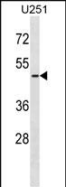
ARMCX3 Antibody (C-term)
Affinity Purified Rabbit Polyclonal Antibody (Pab)
- SPECIFICATION
- CITATIONS
- PROTOCOLS
- BACKGROUND

Application
| WB, E |
|---|---|
| Primary Accession | Q9UH62 |
| Other Accession | NP_057691.1 |
| Reactivity | Human |
| Host | Rabbit |
| Clonality | Polyclonal |
| Isotype | Rabbit IgG |
| Calculated MW | 42501 Da |
| Antigen Region | 299-325 aa |
| Gene ID | 51566 |
|---|---|
| Other Names | Armadillo repeat-containing X-linked protein 3, ARM protein lost in epithelial cancers on chromosome X 3, Protein ALEX3, ARMCX3, ALEX3 |
| Target/Specificity | This ARMCX3 antibody is generated from rabbits immunized with a KLH conjugated synthetic peptide between 299-325 amino acids from the C-terminal region of human ARMCX3. |
| Dilution | WB~~1:1000 E~~Use at an assay dependent concentration. |
| Format | Purified polyclonal antibody supplied in PBS with 0.09% (W/V) sodium azide. This antibody is purified through a protein A column, followed by peptide affinity purification. |
| Storage | Maintain refrigerated at 2-8°C for up to 2 weeks. For long term storage store at -20°C in small aliquots to prevent freeze-thaw cycles. |
| Precautions | ARMCX3 Antibody (C-term) is for research use only and not for use in diagnostic or therapeutic procedures. |
| Name | ARMCX3 |
|---|---|
| Synonyms | ALEX3 |
| Function | Regulates mitochondrial aggregation and transport in axons in living neurons. May link mitochondria to the TRAK2-kinesin motor complex via its interaction with Miro and TRAK2. Mitochondrial distribution and dynamics is regulated through ARMCX3 protein degradation, which is promoted by PCK and negatively regulated by WNT1. Enhances the SOX10-mediated transactivation of the neuronal acetylcholine receptor subunit alpha-3 and beta-4 subunit gene promoters. |
| Cellular Location | Mitochondrion outer membrane {ECO:0000250|UniProtKB:Q8BHS6}; Single-pass membrane protein. Cytoplasm {ECO:0000250|UniProtKB:Q8BHS6}. Nucleus {ECO:0000250|UniProtKB:Q8BHS6} |

Thousands of laboratories across the world have published research that depended on the performance of antibodies from Abcepta to advance their research. Check out links to articles that cite our products in major peer-reviewed journals, organized by research category.
info@abcepta.com, and receive a free "I Love Antibodies" mug.

Provided below are standard protocols that you may find useful for product applications.
Background
This gene encodes a member of the ALEX family of proteins which may play a role in tumor suppression. The encoded protein contains a potential N-terminal transmembrane domain and a single Armadillo (arm) repeat. Other proteins containing the arm repeat are involved in development, maintenance of tissue integrity, and tumorigenesis. This gene is closely localized with other family members on the X chromosome. Three transcript variants encoding the same protein have been identified for this gene. [provided by RefSeq].
References
Mou, Z., et al. J. Biol. Chem. 284(20):13629-13640(2009)
Olsen, J.V., et al. Cell 127(3):635-648(2006)
Olsen, J.V., et al. Cell 127(3):635-648(2006)
Ross, M.T., et al. Nature 434(7031):325-337(2005)
Clark, H.F., et al. Genome Res. 13(10):2265-2270(2003)
If you have used an Abcepta product and would like to share how it has performed, please click on the "Submit Review" button and provide the requested information. Our staff will examine and post your review and contact you if needed.
If you have any additional inquiries please email technical services at tech@abcepta.com.
Ordering Information
Shipping Information
