


 Foundational characteristics of cancer include proliferation, angiogenesis, migration, evasion of apoptosis, and cellular immortality. Find key markers for these cellular processes and antibodies to detect them.
 The SUMOplot™ Analysis Program predicts and scores sumoylation sites in your protein. SUMOylation is a post-translational modification involved in various cellular processes, such as nuclear-cytosolic transport, transcriptional regulation, apoptosis, protein stability, response to stress, and progression through the cell cycle.
 The Autophagy Receptor Motif Plotter predicts and scores autophagy receptor binding sites in your protein. Identifying proteins connected to this pathway is critical to understanding the role of autophagy in physiological as well as pathological processes such as development, differentiation, neurodegenerative diseases, stress, infection, and cancer.
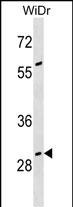
GAS2 Antibody (N-term)
Affinity Purified Rabbit Polyclonal Antibody (Pab)
- SPECIFICATION
- CITATIONS
- PROTOCOLS
- BACKGROUND

Application
| WB, E |
|---|---|
| Primary Accession | O43903 |
| Other Accession | NP_001137302.1 |
| Reactivity | Human |
| Host | Rabbit |
| Clonality | Polyclonal |
| Isotype | Rabbit IgG |
| Calculated MW | 34945 Da |
| Antigen Region | 68-94 aa |
| Gene ID | 2620 |
|---|---|
| Other Names | Growth arrest-specific protein 2, GAS-2, GAS2 |
| Target/Specificity | This GAS2 antibody is generated from rabbits immunized with a KLH conjugated synthetic peptide between 68-94 amino acids from the N-terminal region of human GAS2. |
| Dilution | WB~~1:1000 E~~Use at an assay dependent concentration. |
| Format | Purified polyclonal antibody supplied in PBS with 0.09% (W/V) sodium azide. This antibody is purified through a protein A column, followed by peptide affinity purification. |
| Storage | Maintain refrigerated at 2-8°C for up to 2 weeks. For long term storage store at -20°C in small aliquots to prevent freeze-thaw cycles. |
| Precautions | GAS2 Antibody (N-term) is for research use only and not for use in diagnostic or therapeutic procedures. |
| Name | GAS2 |
|---|---|
| Function | Required to maintain microtubule bundles in inner ear supporting cells, affording them with mechanical stiffness to transmit sound energy through the cochlea. |
| Cellular Location | Cytoplasm, cytoskeleton, stress fiber. Membrane {ECO:0000250|UniProtKB:P11862}; Peripheral membrane protein {ECO:0000250|UniProtKB:P11862} Note=Component of the microfilament system. Colocalizes with actin fibers at the cell border and along the stress fibers in growth- arrested fibroblasts. Mainly membrane-associated. When hyperphosphorylated, accumulates at membrane ruffles (By similarity) Colocalizes with detyrosinated alpha-tubulin along the length of microtubule bundles in inner and outer pillar cells (By similarity) {ECO:0000250|UniProtKB:P11862} |
| Tissue Location | Ubiquitously expressed with highest levels in liver, lung, and kidney. Not found in spleen |

Thousands of laboratories across the world have published research that depended on the performance of antibodies from Abcepta to advance their research. Check out links to articles that cite our products in major peer-reviewed journals, organized by research category.
info@abcepta.com, and receive a free "I Love Antibodies" mug.

Provided below are standard protocols that you may find useful for product applications.
Background
The protein encoded by this gene is a caspase-3 substrate that plays a role in regulating microfilament and cell shape changes during apoptosis. It can also modulate cell susceptibility to p53-dependent apoptosis by inhibiting calpain activity. Multiple alternatively spliced variants, encoding the same protein, have been identified.
References
Huang, W., et al. Mol. Cell. Biol. 30(19):4575-4594(2010)
Shimada, M., et al. Hum. Genet. 128(4):433-441(2010)
Benetti, R., et al. J. Biol. Chem. 280(23):22070-22080(2005)
Tsutsumi, S., et al. Am. J. Hum. Genet. 74(6):1255-1261(2004)
Benetti, R., et al. EMBO J. 20(11):2702-2714(2001)
If you have used an Abcepta product and would like to share how it has performed, please click on the "Submit Review" button and provide the requested information. Our staff will examine and post your review and contact you if needed.
If you have any additional inquiries please email technical services at tech@abcepta.com.
Ordering Information
Other Products
Shipping Information
