


 Foundational characteristics of cancer include proliferation, angiogenesis, migration, evasion of apoptosis, and cellular immortality. Find key markers for these cellular processes and antibodies to detect them.
 The SUMOplot™ Analysis Program predicts and scores sumoylation sites in your protein. SUMOylation is a post-translational modification involved in various cellular processes, such as nuclear-cytosolic transport, transcriptional regulation, apoptosis, protein stability, response to stress, and progression through the cell cycle.
 The Autophagy Receptor Motif Plotter predicts and scores autophagy receptor binding sites in your protein. Identifying proteins connected to this pathway is critical to understanding the role of autophagy in physiological as well as pathological processes such as development, differentiation, neurodegenerative diseases, stress, infection, and cancer.
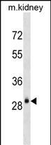
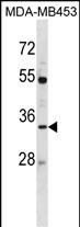
AQP3 Antibody (Center)
Affinity Purified Rabbit Polyclonal Antibody (Pab)
- SPECIFICATION
- CITATIONS
- PROTOCOLS
- BACKGROUND

Application
| WB, E |
|---|---|
| Primary Accession | Q92482 |
| Other Accession | P47862, Q8R2N1, Q08DE6, NP_004916.1 |
| Reactivity | Human, Mouse |
| Predicted | Bovine, Rat |
| Host | Rabbit |
| Clonality | Polyclonal |
| Isotype | Rabbit IgG |
| Calculated MW | 31544 Da |
| Antigen Region | 163-191 aa |
| Gene ID | 360 |
|---|---|
| Other Names | Aquaporin-3, AQP-3, Aquaglyceroporin-3, AQP3 |
| Target/Specificity | This AQP3 antibody is generated from rabbits immunized with a KLH conjugated synthetic peptide between 163-191 amino acids from the Central region of human AQP3. |
| Dilution | WB~~1:1000 E~~Use at an assay dependent concentration. |
| Format | Purified polyclonal antibody supplied in PBS with 0.09% (W/V) sodium azide. This antibody is purified through a protein A column, followed by peptide affinity purification. |
| Storage | Maintain refrigerated at 2-8°C for up to 2 weeks. For long term storage store at -20°C in small aliquots to prevent freeze-thaw cycles. |
| Precautions | AQP3 Antibody (Center) is for research use only and not for use in diagnostic or therapeutic procedures. |
| Name | AQP3 {ECO:0000303|PubMed:7558005, ECO:0000312|HGNC:HGNC:636} |
|---|---|
| Function | Aquaglyceroporins form homotetrameric transmembrane channels, with each monomer independently mediating glycerol and water transport across the plasma membrane along their osmotic gradient (PubMed:12239222, PubMed:30420639). Could also be permeable to urea (By similarity). Also participates in cell permeability to H2O2 and H2O2- mediated signaling (PubMed:20724658). In skin, transports glycerol to the epidermis and stratum corneum, where it maintains hydration, elasticity, and supports lipid biosynthesis for barrier repair (By similarity). In kidney, contributes to the reabsorption of water, helping the body maintain proper fluid balance (By similarity). |
| Cellular Location | Cell membrane; Multi-pass membrane protein {ECO:0000250|UniProtKB:O14520}. Basolateral cell membrane {ECO:0000250|UniProtKB:P47862}; Multi-pass membrane protein {ECO:0000250|UniProtKB:O14520} |
| Tissue Location | Widely expressed in epithelial cells of kidney (collecting ducts) and airways, in keratinocytes, immature dendritic cells and erythrocytes. Isoform 2 is not detectable in erythrocytes at the protein level |

Thousands of laboratories across the world have published research that depended on the performance of antibodies from Abcepta to advance their research. Check out links to articles that cite our products in major peer-reviewed journals, organized by research category.
info@abcepta.com, and receive a free "I Love Antibodies" mug.

Provided below are standard protocols that you may find useful for product applications.
Background
Aquaporin 3 is a water channel protein. Aquaporins are a family of small integral membrane proteins related to the major intrinsic protein (MIP or AQP0). Aquaporin 3 is localized at the basal lateral membranes of collecting duct cells in the kidney. In addition to its water channel function, aquaporin 3 has been found to facilitate the transport of nonionic small solutes such as urea and glycerol, but to a smaller degree. It has been suggested that water channels can be functionally heterogeneous and possess water and solute permeation mechanisms.
References
Bailey, S.D., et al. Diabetes Care 33(10):2250-2253(2010)
Kim, N.H., et al. J. Invest. Dermatol. 130(9):2231-2239(2010)
Ji, C., et al. Int. J. Mol. Med. 26(2):257-263(2010)
Melis, M., et al. Dis. Colon Rectum 53(6):936-943(2010)
Shen, L., et al. Biomed. Pharmacother. 64(5):313-318(2010)
If you have used an Abcepta product and would like to share how it has performed, please click on the "Submit Review" button and provide the requested information. Our staff will examine and post your review and contact you if needed.
If you have any additional inquiries please email technical services at tech@abcepta.com.
Ordering Information
Other Products
Shipping Information
