


 Foundational characteristics of cancer include proliferation, angiogenesis, migration, evasion of apoptosis, and cellular immortality. Find key markers for these cellular processes and antibodies to detect them.
 The SUMOplot™ Analysis Program predicts and scores sumoylation sites in your protein. SUMOylation is a post-translational modification involved in various cellular processes, such as nuclear-cytosolic transport, transcriptional regulation, apoptosis, protein stability, response to stress, and progression through the cell cycle.
 The Autophagy Receptor Motif Plotter predicts and scores autophagy receptor binding sites in your protein. Identifying proteins connected to this pathway is critical to understanding the role of autophagy in physiological as well as pathological processes such as development, differentiation, neurodegenerative diseases, stress, infection, and cancer.
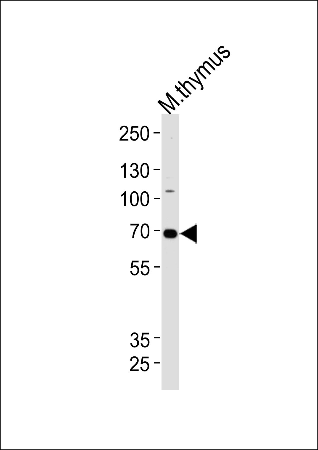
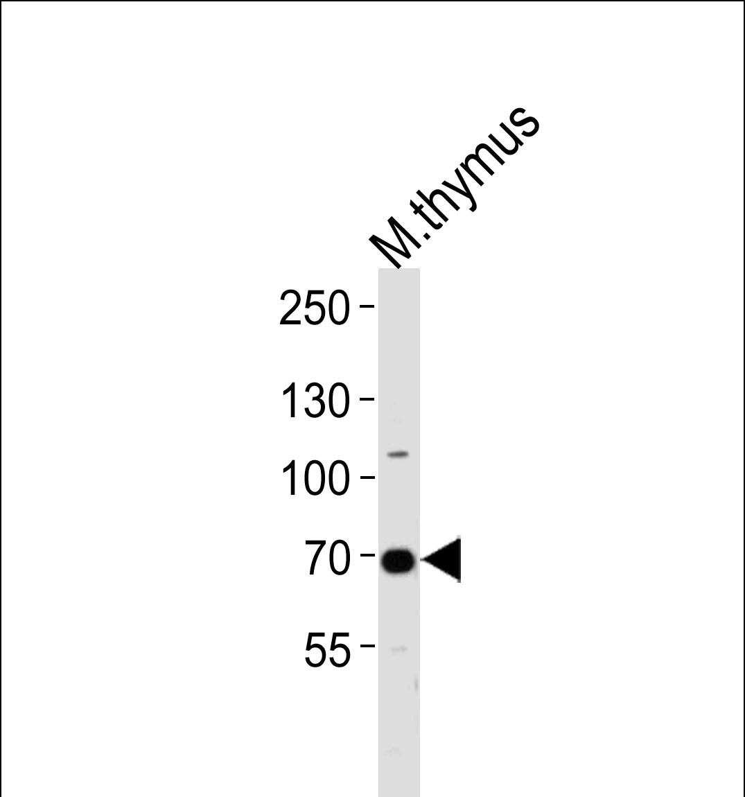
Mouse Ephb6 Antibody(Center)
Affinity Purified Rabbit Polyclonal Antibody (Pab)
- SPECIFICATION
- CITATIONS
- PROTOCOLS
- BACKGROUND

Application
| WB, E |
|---|---|
| Primary Accession | O08644 |
| Other Accession | P0C0K7, O15197, NP_001139823.1 |
| Reactivity | Mouse |
| Predicted | Human, Rat |
| Host | Rabbit |
| Clonality | Polyclonal |
| Isotype | Rabbit IgG |
| Calculated MW | 110107 Da |
| Antigen Region | 503-530 aa |
| Gene ID | 13848 |
|---|---|
| Other Names | Ephrin type-B receptor 6, MEP, Tyrosine-protein kinase-defective receptor EPH-6, Ephb6, Cekl |
| Target/Specificity | This Mouse Ephb6 antibody is generated from rabbits immunized with a KLH conjugated synthetic peptide between 503-530 amino acids from the Central region of mouse Ephb6. |
| Dilution | WB~~1:1000 E~~Use at an assay dependent concentration. |
| Format | Purified polyclonal antibody supplied in PBS with 0.09% (W/V) sodium azide. This antibody is purified through a protein A column, followed by peptide affinity purification. |
| Storage | Maintain refrigerated at 2-8°C for up to 2 weeks. For long term storage store at -20°C in small aliquots to prevent freeze-thaw cycles. |
| Precautions | Mouse Ephb6 Antibody(Center) is for research use only and not for use in diagnostic or therapeutic procedures. |
| Name | Ephb6 |
|---|---|
| Synonyms | Cekl |
| Function | Kinase-defective receptor for members of the ephrin-B family. Binds to ephrin-B1 and ephrin-B2. Modulates cell adhesion and migration by exerting both positive and negative effects upon stimulation with ephrin-B2. Inhibits JNK activation, T-cell receptor-induced IL-2 secretion and CD25 expression upon stimulation with ephrin-B2 (By similarity). |
| Cellular Location | [Isoform 1]: Cell membrane; Single-pass type I membrane protein [Isoform 3]: Secreted. |
| Tissue Location | High level in thymus, and brain. Very low levels of expression in kidney, lung, liver, bone marrow, skeletal muscle, spleen from 2 week old and adult mice, heart, testes and embryonic stem cells |

Thousands of laboratories across the world have published research that depended on the performance of antibodies from Abcepta to advance their research. Check out links to articles that cite our products in major peer-reviewed journals, organized by research category.
info@abcepta.com, and receive a free "I Love Antibodies" mug.

Provided below are standard protocols that you may find useful for product applications.
Background
Kinase-defective receptor for members of the ephrin-B family. Binds to ephrin-B1 and ephrin-B2. Modulates cell adhesion and migration by exerting both positive and negative effects upon stimulation with ephrin-B2. Inhibits JNK activation, T cell receptor-induced IL-2 secretion and CD25 expression upon stimulation with ephrin-B2 (By similarity).
References
Islam, S., et al. Dig. Dis. Sci. 55(9):2478-2488(2010)
Lieben, L., et al. Bone 47(2):301-308(2010)
Mordan-McCombs, S., et al. J. Steroid Biochem. Mol. Biol. 121 (1-2), 368-371 (2010) :
Zirzow, S., et al. Dev. Biol. 336(2):145-155(2009)
Chou, S.J., et al. Nat. Neurosci. 12(11):1381-1389(2009)
If you have used an Abcepta product and would like to share how it has performed, please click on the "Submit Review" button and provide the requested information. Our staff will examine and post your review and contact you if needed.
If you have any additional inquiries please email technical services at tech@abcepta.com.
Ordering Information
Other Products
Shipping Information
