


 Foundational characteristics of cancer include proliferation, angiogenesis, migration, evasion of apoptosis, and cellular immortality. Find key markers for these cellular processes and antibodies to detect them.
 The SUMOplot™ Analysis Program predicts and scores sumoylation sites in your protein. SUMOylation is a post-translational modification involved in various cellular processes, such as nuclear-cytosolic transport, transcriptional regulation, apoptosis, protein stability, response to stress, and progression through the cell cycle.
 The Autophagy Receptor Motif Plotter predicts and scores autophagy receptor binding sites in your protein. Identifying proteins connected to this pathway is critical to understanding the role of autophagy in physiological as well as pathological processes such as development, differentiation, neurodegenerative diseases, stress, infection, and cancer.
PLD3 Antibody(N-term)
Affinity Purified Rabbit Polyclonal Antibody (Pab)
- SPECIFICATION
- CITATIONS
- PROTOCOLS
- BACKGROUND

Application
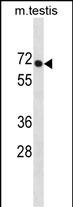
| WB, E |
|---|---|
| Primary Accession | Q8IV08 |
| Other Accession | Q5FVH2, O35405, Q4R583, Q2KJJ8, NP_036400.2 |
| Reactivity | Mouse |
| Predicted | Bovine, Monkey, Rat |
| Host | Rabbit |
| Clonality | Polyclonal |
| Isotype | Rabbit IgG |
| Calculated MW | 54705 Da |
| Antigen Region | 9-37 aa |
| Gene ID | 23646 |
|---|---|
| Other Names | Phospholipase D3, PLD 3, Choline phosphatase 3, HindIII K4L homolog, Hu-K4, Phosphatidylcholine-hydrolyzing phospholipase D3, PLD3 |
| Target/Specificity | This PLD3 antibody is generated from rabbits immunized with a KLH conjugated synthetic peptide between 9-37 amino acids from the N-terminal region of human PLD3. |
| Dilution | WB~~1:1000 E~~Use at an assay dependent concentration. |
| Format | Purified polyclonal antibody supplied in PBS with 0.09% (W/V) sodium azide. This antibody is purified through a protein A column, followed by peptide affinity purification. |
| Storage | Maintain refrigerated at 2-8°C for up to 2 weeks. For long term storage store at -20°C in small aliquots to prevent freeze-thaw cycles. |
| Precautions | PLD3 Antibody(N-term) is for research use only and not for use in diagnostic or therapeutic procedures. |
| Name | PLD3 {ECO:0000303|PubMed:26411346, ECO:0000312|HGNC:HGNC:17158} |
|---|---|
| Function | 5'->3' exonuclease that hydrolyzes the phosphodiester bond of single-stranded DNA (ssDNA) and RNA molecules to form nucleoside 3'- monophosphates and 5'-end 5'-hydroxy deoxyribonucleotide/ribonucleotide fragments (PubMed:30111894, PubMed:30312375, PubMed:34620855, PubMed:37225734, PubMed:37994783, PubMed:38537643, PubMed:38697119). Partially redundant with PLD4, can cleave all four nucleotides displaying higher efficiency for ssDNA and RNA fragments initiated with uridine and guanosine residues and lower efficiency for cytidine- initiated substrates (PubMed:30111894, PubMed:30312375, PubMed:34620855, PubMed:37225734, PubMed:37994783, PubMed:38537643, PubMed:38697119). As a result, it does not always degrade polynucleotides to the single nucleotide level, it can stall at specific sites sparing certain fragments from exonucleolytic degradation (PubMed:30111894, PubMed:30312375, PubMed:34620855, PubMed:37225734, PubMed:37994783, PubMed:38537643, PubMed:38697119). Processes self and pathogenic ssDNA and RNA molecules that reach the endolysosomal compartment via phagocytosis or autophagy and may serve as 'danger' signals for recognition by innate immune receptors such as toll-like receptors (TLRs) (PubMed:34620855, PubMed:37225734, PubMed:38697119). Degrades mitochondrial CpG-rich ssDNA fragments to prevent TLR9 activation and autoinflammatory response, but it can cleave viral RNA to generate ligands for TLR7 activation and initiate antiviral immune responses (PubMed:34620855, PubMed:37225734, PubMed:38697119). In plasmacytoid dendritic cells, it cooperates with endonuclease RNASET2 to release 2',3'-cyclic guanosine monophosphate (2',3'-cGMP), a potent stimulatory ligand for TLR7 (PubMed:34620855, PubMed:37225734, PubMed:38697119). Produces 2',3'-cGMPs and cytidine- rich RNA fragments that occupy TLR7 ligand-binding pockets and trigger a signaling-competent state (PubMed:34620855, PubMed:37225734, PubMed:38697119). Can exert polynucleotide phosphatase activity toward 5'-phosphorylated ssDNA substrates although at a slow rate (PubMed:38537643). Transphosphatidylase that catalyzes the exchange with R to S stereo-inversion of the glycerol moiety between (S,R)- lysophosphatidylglycerol (LPG) and monoacylglycerol (MAG) substrates to yield (S,S)-bis(monoacylglycero)phosphate (BMP) (PubMed:39423811). Can synthesize a variety of (S,S)-BMPs representing the main phospholipid constituent of lysosomal intralumenal vesicle (ILV) membranes that bind acid hydrolases for lipid degradation (PubMed:39423811). Regulates the homeostasis and interorganellar communication of the endolysosomal system with an overall impact on cellular removal of dysfunctional organelles via autophagy as well as proper protein and lipid turnover (PubMed:28128235, PubMed:29368044, PubMed:37225734). May play a role in myotube formation in response to ER stress (PubMed:22428023). |
| Cellular Location | Endoplasmic reticulum membrane; Single-pass type II membrane protein. Lysosome lumen. Early endosome membrane; Single-pass type II membrane protein. Late endosome membrane; Single-pass type II membrane protein. Golgi apparatus membrane; Single-pass type II membrane protein. Endosome membrane; Single-pass type II membrane protein. Note=Localizes to ER-associated vesicles in differentiating myotubes (PubMed:22428023). Sorted into intralumenal vesicles (ILVs) in lysosomes. The soluble form in lysosome arises by proteolytic processing of the membrane-bound form (PubMed:29386126) Colocalizes with APP in endosomes (PubMed:29368044) |
| Tissue Location | Widely expressed. In the brain, high levels of expression are detected in the frontal, temporal and occipital cortices and hippocampus. Expressed at low level in corpus callosum. Expressed in plasmacytoid dendritic cells and monocytes (at protein level) |

Thousands of laboratories across the world have published research that depended on the performance of antibodies from Abcepta to advance their research. Check out links to articles that cite our products in major peer-reviewed journals, organized by research category.
info@abcepta.com, and receive a free "I Love Antibodies" mug.

Provided below are standard protocols that you may find useful for product applications.
Background
The function of this protein is unknown.
References
Idkowiak-Baldys, J., et al. J. Biol. Chem. 284(33):22322-22331(2009)
Walker, L.C., et al. Breast Cancer Res. Treat. 112(2):229-236(2008)
Russo, P., et al. Am. J. Hypertens. 19(4):331-338(2006)
Cheng, J., et al. Science 308(5725):1149-1154(2005)
Munck, A., et al. FEBS J. 272(7):1718-1726(2005)
If you have used an Abcepta product and would like to share how it has performed, please click on the "Submit Review" button and provide the requested information. Our staff will examine and post your review and contact you if needed.
If you have any additional inquiries please email technical services at tech@abcepta.com.
Ordering Information
Other Products
Shipping Information
