


 Foundational characteristics of cancer include proliferation, angiogenesis, migration, evasion of apoptosis, and cellular immortality. Find key markers for these cellular processes and antibodies to detect them.
 The SUMOplot™ Analysis Program predicts and scores sumoylation sites in your protein. SUMOylation is a post-translational modification involved in various cellular processes, such as nuclear-cytosolic transport, transcriptional regulation, apoptosis, protein stability, response to stress, and progression through the cell cycle.
 The Autophagy Receptor Motif Plotter predicts and scores autophagy receptor binding sites in your protein. Identifying proteins connected to this pathway is critical to understanding the role of autophagy in physiological as well as pathological processes such as development, differentiation, neurodegenerative diseases, stress, infection, and cancer.
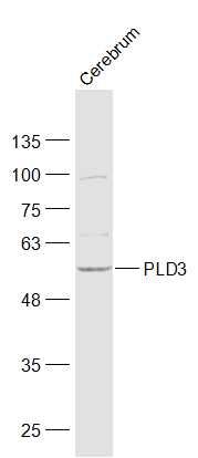
PLD3 Polyclonal Antibody
Purified Rabbit Polyclonal Antibody (Pab)
- SPECIFICATION
- CITATIONS
- PROTOCOLS
- BACKGROUND

Application
| WB, IHC-P, IHC-F, IF, ICC |
|---|---|
| Primary Accession | Q8IV08 |
| Reactivity | Rat, Bovine |
| Host | Rabbit |
| Clonality | Polyclonal |
| Calculated MW | 54705 Da |
| Gene ID | 23646 |
|---|---|
| Other Names | 5'-3' exonuclease PLD3, 3.1.16.1, Choline phosphatase 3, HindIII K4L homolog, Hu-K4, Phosphatidylcholine-hydrolyzing phospholipase D3, Phospholipase D3, PLD 3, PLD3 |
| Dilution | WB=1:500-2000,Elisa=1:500-1000,IHC-P=1:100-500,IHC-F=1:100-500,IF=1:100-500,ICC=1:100-500, |
| Format | 0.01M TBS(pH7.4), 0.09% (W/V) sodium azide and 50% Glyce |
| Storage | Store at -20 ℃ for one year. Avoid repeated freeze/thaw cycles. When reconstituted in sterile pH 7.4 0.01M PBS or diluent of antibody the antibody is stable for at least two weeks at 2-4 ℃. |
| Name | PLD3 |
|---|---|
| Function | 5'->3' DNA exonuclease which digests single-stranded DNA (ssDNA) (PubMed:30312375). Regulates inflammatory cytokine responses via the degradation of nucleic acids, by reducing the concentration of ssDNA able to stimulate TLR9, a nucleotide-sensing receptor in collaboration with PLD4 (By similarity). May be important in myotube formation (PubMed:22428023). Plays a role in lysosomal homeostasis (PubMed:28128235). Involved in the regulation of endosomal protein sorting (PubMed:29368044). |
| Cellular Location | Endoplasmic reticulum membrane; Single-pass type II membrane protein. Lysosome lumen. Early endosome membrane; Single-pass type II membrane protein. Late endosome membrane; Single-pass type II membrane protein. Golgi apparatus membrane; Single-pass type II membrane protein. Endosome membrane; Single-pass type II membrane protein. Note=Localizes to ER-associated vesicles in differentiating myotubes (PubMed:22428023). The soluble form in lysosome arises by proteolytic processing of the membrane-bound form (PubMed:29386126). Colocalizes with APP in endosomes (PubMed:29368044) |
| Tissue Location | Widely expressed. In the brain, high levels of expression are detected in the frontal, temporal and occipital cortices and hippocampus. Expressed at low level in corpus callosum |
Research Areas
Citations (0)

Thousands of laboratories across the world have published research that depended on the performance of antibodies from Abcepta to advance their research. Check out links to articles that cite our products in major peer-reviewed journals, organized by research category.
Submit your citation using an Abcepta antibody to
info@abcepta.com, and receive a free "I Love Antibodies" mug.
info@abcepta.com, and receive a free "I Love Antibodies" mug.

Application Protocols
Provided below are standard protocols that you may find useful for product applications.
Abcepta welcomes feedback from its customers.
If you have used an Abcepta product and would like to share how it has performed, please click on the "Submit Review" button and provide the requested information. Our staff will examine and post your review and contact you if needed.
If you have any additional inquiries please email technical services at tech@abcepta.com.
$ 350.00
Cat# AP54943
Ordering Information
United States
AlbaniaAustraliaAustriaBelgiumBosnia & HerzegovinaBrazilBulgariaCanadaCentral AmericaChinaCroatiaCyprusCzech RepublicDenmarkEstoniaFinlandFranceGermanyGreeceHong KongHungaryIcelandIndiaIndonesiaIrelandIsraelItalyJapanLatviaLithuaniaLuxembourgMacedoniaMalaysiaMaltaNetherlandsNew ZealandNorwayPakistanPolandPortugalRomaniaSerbiaSingaporeSlovakiaSloveniaSouth AfricaSouth KoreaSpainSwedenSwitzerlandTaiwanTurkeyUnited KingdomUnited StatesVietnamWorldwideOthers
USA Headquarters
(888) 735-7227 / (858) 622-0099 or (858) 875-1900
Other Products
Shipping Information
Domestic orders (in stock items)
Shipped out the same day. Orders placed after 1 PM (PST) will ship out the next business day.
International orders
Contact your local distributors
