


 Foundational characteristics of cancer include proliferation, angiogenesis, migration, evasion of apoptosis, and cellular immortality. Find key markers for these cellular processes and antibodies to detect them.
 The SUMOplot™ Analysis Program predicts and scores sumoylation sites in your protein. SUMOylation is a post-translational modification involved in various cellular processes, such as nuclear-cytosolic transport, transcriptional regulation, apoptosis, protein stability, response to stress, and progression through the cell cycle.
 The Autophagy Receptor Motif Plotter predicts and scores autophagy receptor binding sites in your protein. Identifying proteins connected to this pathway is critical to understanding the role of autophagy in physiological as well as pathological processes such as development, differentiation, neurodegenerative diseases, stress, infection, and cancer.
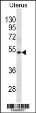
KRT72 Antibody(Center)
Affinity Purified Rabbit Polyclonal Antibody (Pab)
- SPECIFICATION
- CITATIONS
- PROTOCOLS
- BACKGROUND

Application
| WB, E |
|---|---|
| Primary Accession | Q14CN4 |
| Other Accession | Q148H8, NP_542785.1 |
| Reactivity | Human |
| Predicted | Bovine |
| Host | Rabbit |
| Clonality | Polyclonal |
| Isotype | Rabbit IgG |
| Calculated MW | 55877 Da |
| Antigen Region | 313-342 aa |
| Gene ID | 140807 |
|---|---|
| Other Names | Keratin, type II cytoskeletal 72, Cytokeratin-72, CK-72, Keratin-72, K72, Type II inner root sheath-specific keratin-K6irs2, Type-II keratin Kb35, KRT72, K6IRS2, KB35, KRT6, KRT6IRS2 |
| Target/Specificity | This KRT72 antibody is generated from rabbits immunized with a KLH conjugated synthetic peptide between 313-342 amino acids from the Central region of human KRT72. |
| Dilution | WB~~1:1000 E~~Use at an assay dependent concentration. |
| Format | Purified polyclonal antibody supplied in PBS with 0.09% (W/V) sodium azide. This antibody is purified through a protein A column, followed by peptide affinity purification. |
| Storage | Maintain refrigerated at 2-8°C for up to 2 weeks. For long term storage store at -20°C in small aliquots to prevent freeze-thaw cycles. |
| Precautions | KRT72 Antibody(Center) is for research use only and not for use in diagnostic or therapeutic procedures. |
| Name | KRT72 |
|---|---|
| Synonyms | K6IRS2, KB35, KRT6, KRT6IRS2 |
| Function | Has a role in hair formation. Specific component of keratin intermediate filaments in the inner root sheath (IRS) of the hair follicle (Probable). |
| Tissue Location | Highly expressed in hair follicles from scalp and eyebrow. Also expressed in palmoplantar epidermis. Not expressed in face skin despite the presence of fine hairs histologically. In hair, it is specifically present in the inner root sheath (IRS) of the hair follicle. Present in the IRS cuticle, but not in Henle or Huxley layers of the IRS. In the IRS cuticle, its presence is delayed up to the height of the apex of the dermal papilla (at protein level) |

Thousands of laboratories across the world have published research that depended on the performance of antibodies from Abcepta to advance their research. Check out links to articles that cite our products in major peer-reviewed journals, organized by research category.
info@abcepta.com, and receive a free "I Love Antibodies" mug.

Provided below are standard protocols that you may find useful for product applications.
Background
Keratins are intermediate filament proteins responsible for the structural integrity of epithelial cells. The type II keratins consist of basic or neutral proteins which are arranged in pairs of heterotypic keratin chains coexpressed during differentiation of simple and stratified epithelial tissues. This gene encodes a type II keratin that is specifically expressed in the inner root sheath of hair follicles. The type II keratins are clustered in a region of chromosome 12q12-q13. Multiple transcript variants encoding different isoforms have been found for this gene.
References
Bertheau, P., et al. PLoS Med. 4 (3), E90 (2007) :
Schweizer, J., et al. J. Cell Biol. 174(2):169-174(2006)
Langbein, L., et al. J. Invest. Dermatol. 120(4):512-522(2003)
Porter, R.M., et al. Br. J. Dermatol. 145(4):558-568(2001)
If you have used an Abcepta product and would like to share how it has performed, please click on the "Submit Review" button and provide the requested information. Our staff will examine and post your review and contact you if needed.
If you have any additional inquiries please email technical services at tech@abcepta.com.
Ordering Information
Other Products
Shipping Information
