


 Foundational characteristics of cancer include proliferation, angiogenesis, migration, evasion of apoptosis, and cellular immortality. Find key markers for these cellular processes and antibodies to detect them.
 The SUMOplot™ Analysis Program predicts and scores sumoylation sites in your protein. SUMOylation is a post-translational modification involved in various cellular processes, such as nuclear-cytosolic transport, transcriptional regulation, apoptosis, protein stability, response to stress, and progression through the cell cycle.
 The Autophagy Receptor Motif Plotter predicts and scores autophagy receptor binding sites in your protein. Identifying proteins connected to this pathway is critical to understanding the role of autophagy in physiological as well as pathological processes such as development, differentiation, neurodegenerative diseases, stress, infection, and cancer.
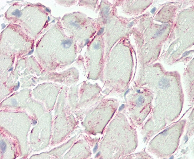
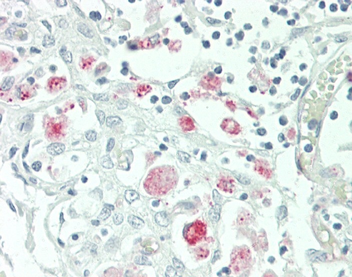
TMIGD2 Antibody (Center)
Affinity Purified Rabbit Polyclonal Antibody (Pab)

- SPECIFICATION
- CITATIONS
- PROTOCOLS
- BACKGROUND

Application
| WB, IHC-P, E |
|---|---|
| Primary Accession | Q96BF3 |
| Other Accession | NP_653216.2 |
| Reactivity | Human |
| Host | Rabbit |
| Clonality | Polyclonal |
| Isotype | Rabbit IgG |
| Calculated MW | 30675 Da |
| Antigen Region | 119-147 aa |
| Gene ID | 126259 |
|---|---|
| Other Names | Transmembrane and immunoglobulin domain-containing protein 2, CD28 homolog, Immunoglobulin and proline-rich receptor 1, IGPR-1, TMIGD2, CD28H, IGPR1 |
| Target/Specificity | This TMIGD2 antibody is generated from rabbits immunized with a KLH conjugated synthetic peptide between 119-147 amino acids from the Central region of human TMIGD2. |
| Dilution | WB~~1:1000 IHC-P~~1:100 E~~Use at an assay dependent concentration. |
| Format | Purified polyclonal antibody supplied in PBS with 0.09% (W/V) sodium azide. This antibody is purified through a protein A column, followed by peptide affinity purification. |
| Storage | Maintain refrigerated at 2-8°C for up to 2 weeks. For long term storage store at -20°C in small aliquots to prevent freeze-thaw cycles. |
| Precautions | TMIGD2 Antibody (Center) is for research use only and not for use in diagnostic or therapeutic procedures. |
| Name | TMIGD2 |
|---|---|
| Synonyms | CD28H, IGPR1 |
| Function | Plays a role in cell-cell interaction, cell migration, and angiogenesis. Through interaction with HHLA2, costimulates T-cells in the context of TCR-mediated activation. Enhances T-cell proliferation and cytokine production via an AKT-dependent signaling cascade. |
| Cellular Location | Cell membrane; Single-pass type I membrane protein |
| Tissue Location | Widely expressed, mainly by epithelial and endothelial cells, including bronchial epithelial cells of lung, breast glandular and lobular epithelia cells, urothelium of the bladder, skin epidermis, epithelium of gastrointestinal, rectum, endometrial glands of the uterus, ureter, fallopian tube epithelium, colonic epithelium, small bowl epithelium, stomach epithelium, including both chief and parietal cells, trophoblastic epithelium of placenta, and pancreatic acinar cells (at protein level). Consistently expressed in veins and arteries (at protein level). Not detected in thyroid, cerebellum, cerebral cortex and thymus (at protein level). Expressed in lymphoid organs, with highest levels in thymus, spleen, peripheral blood lymphocytes and liver. In the thymus, expressed in CD4+ and CD8+ single- and double-positive cells, but not in immature CD4- and CD8- double-negative cells (at protein level). In peripheral blood mononuclear cells, highly expressed on CD56+ or CD16+ natural killer cells and CD3+ T-cells(at protein level). Not detected on B-cells(at protein level). Expressed in tonsils (at protein level) |

Thousands of laboratories across the world have published research that depended on the performance of antibodies from Abcepta to advance their research. Check out links to articles that cite our products in major peer-reviewed journals, organized by research category.
info@abcepta.com, and receive a free "I Love Antibodies" mug.

Provided below are standard protocols that you may find useful for product applications.
Background
The function of this protein is unknown.
References
Gerhard, D.S., et al. Genome Res. 14 (10B), 2121-2127 (2004) :
If you have used an Abcepta product and would like to share how it has performed, please click on the "Submit Review" button and provide the requested information. Our staff will examine and post your review and contact you if needed.
If you have any additional inquiries please email technical services at tech@abcepta.com.
Ordering Information
Other Products
Shipping Information
