


 Foundational characteristics of cancer include proliferation, angiogenesis, migration, evasion of apoptosis, and cellular immortality. Find key markers for these cellular processes and antibodies to detect them.
 The SUMOplot™ Analysis Program predicts and scores sumoylation sites in your protein. SUMOylation is a post-translational modification involved in various cellular processes, such as nuclear-cytosolic transport, transcriptional regulation, apoptosis, protein stability, response to stress, and progression through the cell cycle.
 The Autophagy Receptor Motif Plotter predicts and scores autophagy receptor binding sites in your protein. Identifying proteins connected to this pathway is critical to understanding the role of autophagy in physiological as well as pathological processes such as development, differentiation, neurodegenerative diseases, stress, infection, and cancer.
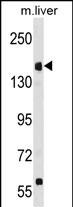
UBR1 Antibody (N-term)
Affinity Purified Rabbit Polyclonal Antibody (Pab)
- SPECIFICATION
- CITATIONS
- PROTOCOLS
- BACKGROUND

Application
| WB, E |
|---|---|
| Primary Accession | Q8IWV7 |
| Other Accession | O70481, NP_777576.1 |
| Reactivity | Mouse |
| Host | Rabbit |
| Clonality | Polyclonal |
| Isotype | Rabbit IgG |
| Calculated MW | 200211 Da |
| Antigen Region | 245-274 aa |
| Gene ID | 197131 |
|---|---|
| Other Names | E3 ubiquitin-protein ligase UBR1, 632-, N-recognin-1, Ubiquitin-protein ligase E3-alpha-1, Ubiquitin-protein ligase E3-alpha-I, UBR1 |
| Target/Specificity | This UBR1 antibody is generated from rabbits immunized with a KLH conjugated synthetic peptide between 245-274 amino acids from the N-terminal region of human UBR1. |
| Dilution | WB~~1:1000 E~~Use at an assay dependent concentration. |
| Format | Purified polyclonal antibody supplied in PBS with 0.09% (W/V) sodium azide. This antibody is purified through a protein A column, followed by peptide affinity purification. |
| Storage | Maintain refrigerated at 2-8°C for up to 2 weeks. For long term storage store at -20°C in small aliquots to prevent freeze-thaw cycles. |
| Precautions | UBR1 Antibody (N-term) is for research use only and not for use in diagnostic or therapeutic procedures. |
| Name | UBR1 {ECO:0000303|PubMed:15317757, ECO:0000312|HGNC:HGNC:16808} |
|---|---|
| Function | E3 ubiquitin-protein ligase which is a component of the N-end rule pathway (PubMed:15548684, PubMed:16311597, PubMed:18162545, PubMed:20835242, PubMed:28392261). Recognizes and binds proteins bearing specific N-terminal residues that are destabilizing according to the N-end rule, leading to their ubiquitination and subsequent degradation (PubMed:18162545, PubMed:20835242, PubMed:28392261). Recognizes both type-1 and type-2 N-degrons, containing positively charged amino acids (Arg, Lys and His) and bulky and hydrophobic amino acids, respectively (PubMed:18162545). Does not ubiquitinate proteins that are acetylated at the N-terminus (PubMed:20835242). In contrast, it strongly binds methylated N-degrons (PubMed:28392261). Binds leucine and is a negative regulator of the leucine-mTOR signaling pathway, thereby controlling cell growth (PubMed:20298436). |
| Cellular Location | Cytoplasm, cytosol. |
| Tissue Location | Broadly expressed, with highest levels in skeletal muscle, kidney and pancreas. Present in acinar cells of the pancreas (at protein level). |

Thousands of laboratories across the world have published research that depended on the performance of antibodies from Abcepta to advance their research. Check out links to articles that cite our products in major peer-reviewed journals, organized by research category.
info@abcepta.com, and receive a free "I Love Antibodies" mug.

Provided below are standard protocols that you may find useful for product applications.
Background
The N-end rule pathway is one proteolytic pathway of the ubiquitin system. The recognition component of this pathway, encoded by this gene, binds to a destabilizing N-terminal residue of a substrate protein and participates in the formation of a substrate-linked multiubiquitin chain. This leads to the eventual degradation of the substrate protein. The protein described in this record has a RING-type zinc finger and a UBR-type zinc finger. Mutations in this gene have been associated with Johanson-Blizzard syndrome.
References
Elting, M., et al. Am. J. Med. Genet. A 146A (23), 3058-3061 (2008) :
Alkhouri, N., et al. World J. Gastroenterol. 14(44):6863-6866(2008)
Yamaguchi, Y., et al. BMC Med. Genet. 9, 22 (2008) :
Schmidt, R.L., et al. Cancer Res. 67(24):11798-11810(2007)
Wei, S., et al. Mol. Pharmacol. 72(3):725-733(2007)
If you have used an Abcepta product and would like to share how it has performed, please click on the "Submit Review" button and provide the requested information. Our staff will examine and post your review and contact you if needed.
If you have any additional inquiries please email technical services at tech@abcepta.com.
Ordering Information
Other Products
Shipping Information
