


 Foundational characteristics of cancer include proliferation, angiogenesis, migration, evasion of apoptosis, and cellular immortality. Find key markers for these cellular processes and antibodies to detect them.
 The SUMOplot™ Analysis Program predicts and scores sumoylation sites in your protein. SUMOylation is a post-translational modification involved in various cellular processes, such as nuclear-cytosolic transport, transcriptional regulation, apoptosis, protein stability, response to stress, and progression through the cell cycle.
 The Autophagy Receptor Motif Plotter predicts and scores autophagy receptor binding sites in your protein. Identifying proteins connected to this pathway is critical to understanding the role of autophagy in physiological as well as pathological processes such as development, differentiation, neurodegenerative diseases, stress, infection, and cancer.
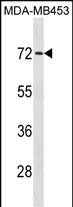
MEX3C Antibody (C-term)
Affinity Purified Rabbit Polyclonal Antibody (Pab)
- SPECIFICATION
- CITATIONS
- PROTOCOLS
- BACKGROUND

Application
| WB, E |
|---|---|
| Primary Accession | Q5U5Q3 |
| Other Accession | Q05A36, NP_057710.3 |
| Reactivity | Human |
| Predicted | Mouse |
| Host | Rabbit |
| Clonality | Polyclonal |
| Isotype | Rabbit IgG |
| Calculated MW | 69366 Da |
| Antigen Region | 536-564 aa |
| Gene ID | 51320 |
|---|---|
| Other Names | RNA-binding E3 ubiquitin-protein ligase MEX3C, 632-, RING finger and KH domain-containing protein 2, RING finger protein 194, MEX3C, RKHD2, RNF194 |
| Target/Specificity | This MEX3C antibody is generated from rabbits immunized with a KLH conjugated synthetic peptide between 536-564 amino acids from the C-terminal region of human MEX3C. |
| Dilution | WB~~1:1000 E~~Use at an assay dependent concentration. |
| Format | Purified polyclonal antibody supplied in PBS with 0.09% (W/V) sodium azide. This antibody is purified through a protein A column, followed by peptide affinity purification. |
| Storage | Maintain refrigerated at 2-8°C for up to 2 weeks. For long term storage store at -20°C in small aliquots to prevent freeze-thaw cycles. |
| Precautions | MEX3C Antibody (C-term) is for research use only and not for use in diagnostic or therapeutic procedures. |
| Name | MEX3C |
|---|---|
| Synonyms | RKHD2, RNF194 |
| Function | E3 ubiquitin ligase responsible for the post-transcriptional regulation of common HLA-A allotypes. Binds to the 3' UTR of HLA-A2 mRNA, and regulates its levels by promoting mRNA decay. RNA binding is sufficient to prevent translation, but ubiquitin ligase activity is required for mRNA degradation. |
| Cellular Location | Cytoplasm. Nucleus. Note=Predominantly expressed in the cytoplasm and shuttles between the cytoplasm and the nucleus through the CRM1 export pathway. May act as suppressor of replication stress and chromosome missegregation |
| Tissue Location | Highest levels found in fetal brain and testis. Also expressed in thymus, salivary gland and uterus. Highly expressed in cells of the innate immune system, in particular activated NK cells Week expression in the intestine. |

Thousands of laboratories across the world have published research that depended on the performance of antibodies from Abcepta to advance their research. Check out links to articles that cite our products in major peer-reviewed journals, organized by research category.
info@abcepta.com, and receive a free "I Love Antibodies" mug.

Provided below are standard protocols that you may find useful for product applications.
Background
This gene encodes a member of a family of proteins with two K homology (KH) RNA-binding domains and a C-terminal RING-finger domain. The protein interacts with mRNA via the KH domains, and the protein shuttles between the nucleus and cytoplasm. Polymorphisms in this gene may contribute to hypertension.
References
Rose, J.E., et al. Mol. Med. 16 (7-8), 247-253 (2010) :
Buchet-Poyau, K., et al. Nucleic Acids Res. 35(4):1289-1300(2007)
Guzman, B., et al. Hypertension 48(5):883-891(2006)
Nusbaum, C., et al. Nature 437(7058):551-555(2005)
If you have used an Abcepta product and would like to share how it has performed, please click on the "Submit Review" button and provide the requested information. Our staff will examine and post your review and contact you if needed.
If you have any additional inquiries please email technical services at tech@abcepta.com.
Ordering Information
Other Products
Shipping Information
