


 Foundational characteristics of cancer include proliferation, angiogenesis, migration, evasion of apoptosis, and cellular immortality. Find key markers for these cellular processes and antibodies to detect them.
 The SUMOplot™ Analysis Program predicts and scores sumoylation sites in your protein. SUMOylation is a post-translational modification involved in various cellular processes, such as nuclear-cytosolic transport, transcriptional regulation, apoptosis, protein stability, response to stress, and progression through the cell cycle.
 The Autophagy Receptor Motif Plotter predicts and scores autophagy receptor binding sites in your protein. Identifying proteins connected to this pathway is critical to understanding the role of autophagy in physiological as well as pathological processes such as development, differentiation, neurodegenerative diseases, stress, infection, and cancer.
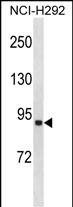
ACSL1 Antibody (C-term)
Affinity Purified Rabbit Polyclonal Antibody (Pab)
- SPECIFICATION
- CITATIONS
- PROTOCOLS
- BACKGROUND

Application
| WB, E |
|---|---|
| Primary Accession | P33121 |
| Other Accession | NP_001986.2 |
| Reactivity | Human |
| Host | Rabbit |
| Clonality | Polyclonal |
| Isotype | Rabbit IgG |
| Calculated MW | 77943 Da |
| Antigen Region | 505-533 aa |
| Gene ID | 2180 |
|---|---|
| Other Names | Long-chain-fatty-acid--CoA ligase 1, Acyl-CoA synthetase 1, ACS1, Long-chain acyl-CoA synthetase 1, LACS 1, Long-chain acyl-CoA synthetase 2, LACS 2, Long-chain fatty acid-CoA ligase 2, Palmitoyl-CoA ligase 1, Palmitoyl-CoA ligase 2, ACSL1, FACL1, FACL2, LACS, LACS1, LACS2 |
| Target/Specificity | This ACSL1 antibody is generated from rabbits immunized with a KLH conjugated synthetic peptide between 505-533 amino acids from the C-terminal region of human ACSL1. |
| Dilution | WB~~1:1000 E~~Use at an assay dependent concentration. |
| Format | Purified polyclonal antibody supplied in PBS with 0.09% (W/V) sodium azide. This antibody is purified through a protein A column, followed by peptide affinity purification. |
| Storage | Maintain refrigerated at 2-8°C for up to 2 weeks. For long term storage store at -20°C in small aliquots to prevent freeze-thaw cycles. |
| Precautions | ACSL1 Antibody (C-term) is for research use only and not for use in diagnostic or therapeutic procedures. |
| Name | ACSL1 (HGNC:3569) |
|---|---|
| Function | Catalyzes the conversion of long-chain fatty acids to their active form acyl-CoAs for both synthesis of cellular lipids, and degradation via beta-oxidation (PubMed:21242590, PubMed:22633490, PubMed:24269233). Preferentially uses palmitoleate, oleate and linoleate (PubMed:24269233). Preferentially activates arachidonate than epoxyeicosatrienoic acids (EETs) or hydroxyeicosatrienoic acids (HETEs) (By similarity). |
| Cellular Location | Mitochondrion outer membrane; Single-pass type III membrane protein. Peroxisome membrane; Single-pass type III membrane protein. Microsome membrane; Single-pass type III membrane protein. Endoplasmic reticulum membrane; Single-pass type III membrane protein |
| Tissue Location | Highly expressed in liver, heart, skeletal muscle, kidney and erythroid cells, and to a lesser extent in brain, lung, placenta and pancreas. |

Thousands of laboratories across the world have published research that depended on the performance of antibodies from Abcepta to advance their research. Check out links to articles that cite our products in major peer-reviewed journals, organized by research category.
info@abcepta.com, and receive a free "I Love Antibodies" mug.

Provided below are standard protocols that you may find useful for product applications.
Background
The protein encoded by this gene is an isozyme of the long-chain fatty-acid-coenzyme A ligase family. Although differing in substrate specificity, subcellular localization, and tissue distribution, all isozymes of this family convert free long-chain fatty acids into fatty acyl-CoA esters, and thereby play a key role in lipid biosynthesis and fatty acid degradation. [provided by RefSeq].
References
Phillips, C.M., et al. J. Lipid Res. 51(7):1793-1800(2010)
Lu, Y., et al. J. Lipid Res. 49(12):2582-2589(2008)
Soupene, E., et al. BMC Mol. Biol. 7, 21 (2006) :
Kahn, B.B., et al. Cell Metab. 1(1):15-25(2005)
Mashek, D.G., et al. J. Lipid Res. 45(10):1958-1961(2004)
If you have used an Abcepta product and would like to share how it has performed, please click on the "Submit Review" button and provide the requested information. Our staff will examine and post your review and contact you if needed.
If you have any additional inquiries please email technical services at tech@abcepta.com.
Ordering Information
Other Products
Shipping Information
