


 Foundational characteristics of cancer include proliferation, angiogenesis, migration, evasion of apoptosis, and cellular immortality. Find key markers for these cellular processes and antibodies to detect them.
 The SUMOplot™ Analysis Program predicts and scores sumoylation sites in your protein. SUMOylation is a post-translational modification involved in various cellular processes, such as nuclear-cytosolic transport, transcriptional regulation, apoptosis, protein stability, response to stress, and progression through the cell cycle.
 The Autophagy Receptor Motif Plotter predicts and scores autophagy receptor binding sites in your protein. Identifying proteins connected to this pathway is critical to understanding the role of autophagy in physiological as well as pathological processes such as development, differentiation, neurodegenerative diseases, stress, infection, and cancer.
ACSL1 Antibody
- SPECIFICATION
- CITATIONS
- PROTOCOLS
- BACKGROUND

Application
| WB, IF, E |
|---|---|
| Primary Accession | P33121 |
| Other Accession | NP_001986, 40807491 |
| Reactivity | Human, Mouse, Rat |
| Host | Rabbit |
| Clonality | Polyclonal |
| Isotype | IgG |
| Calculated MW | 77 kDa |
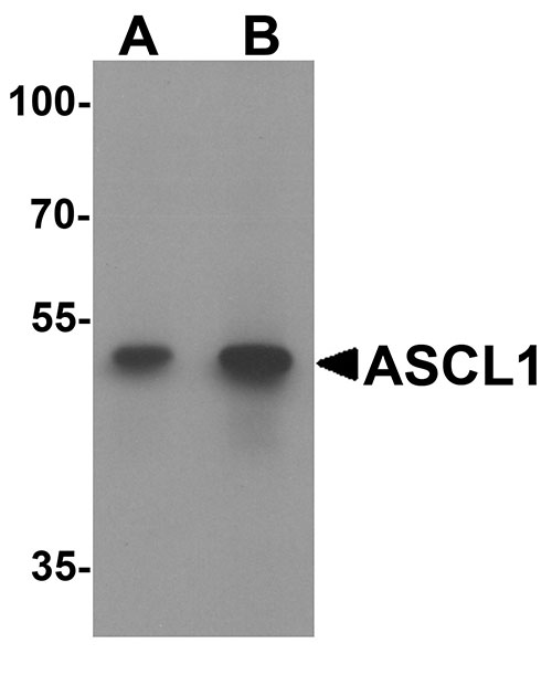
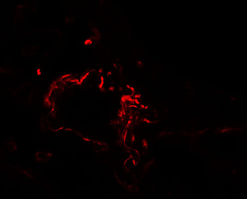
| Application Notes | ACSL1 antibody can be used for detection of ACSL1 by Western blot at 1 - 2 µg/mL. For immunofluorescence start at 20 µg/mL. |
| Gene ID | 2180 |
|---|---|
| Target/Specificity | ACSL1; At least three isoforms of ACSL1 are known to exist; this antibody will detect all three isoforms. |
| Reconstitution & Storage | ACSL1 antibody can be stored at 4℃ for three months and -20℃, stable for up to one year. As with all antibodies care should be taken to avoid repeated freeze thaw cycles. Antibodies should not be exposed to prolonged high temperatures. |
| Precautions | ACSL1 Antibody is for research use only and not for use in diagnostic or therapeutic procedures. |
| Name | ACSL1 (HGNC:3569) |
|---|---|
| Function | Catalyzes the conversion of long-chain fatty acids to their active form acyl-CoAs for both synthesis of cellular lipids, and degradation via beta-oxidation (PubMed:21242590, PubMed:22633490, PubMed:24269233). Preferentially uses palmitoleate, oleate and linoleate (PubMed:24269233). Preferentially activates arachidonate than epoxyeicosatrienoic acids (EETs) or hydroxyeicosatrienoic acids (HETEs) (By similarity). |
| Cellular Location | Mitochondrion outer membrane; Single-pass type III membrane protein. Peroxisome membrane; Single-pass type III membrane protein. Microsome membrane; Single-pass type III membrane protein. Endoplasmic reticulum membrane; Single-pass type III membrane protein |
| Tissue Location | Highly expressed in liver, heart, skeletal muscle, kidney and erythroid cells, and to a lesser extent in brain, lung, placenta and pancreas. |

Thousands of laboratories across the world have published research that depended on the performance of antibodies from Abcepta to advance their research. Check out links to articles that cite our products in major peer-reviewed journals, organized by research category.
info@abcepta.com, and receive a free "I Love Antibodies" mug.

Provided below are standard protocols that you may find useful for product applications.
Background
ACSL1 Antibody: Long-chain acyl coenzyme A synthetase 1 (ACSL1) catalyzes the synthesis of acyl-CoA from long-chain fatty acids in an ATP-dependent manner. ACSL1 is a member of a family of long-chain acyl-CoA synthetases which differ in substrate preference, tissue expression, and subcellular localization. In mouse, ASCL1 is the major acyl-CoA enzyme in the heart, providing 60-90% of heart ATP. Loss of ASCL1 either globally or in heart ventricles resulted in impaired fatty acid oxidation, activation of the mammalian target of rapamycin (mTOR), and cardiac hypertrophy.
References
Black PN and DiRusso CC. Transmembrane movement of exogenous long-chain fatty acids: proteins, enzymes, and vectorial esterification. Microbiol. Mol. Biol. Rev. 2003; 67:454-72.
Coleman RA, Lewin TM, Van Horn CG, et al. Do acyl-CoA synthetases regulate fatty acid entry into synthetic versus degradative pathways? J. Nutr. 2002; 132:2123-6.
Clark H, Carling D, and Saggerson D. Covalent activation of heart AMP-activated protein kinase in response to physiological concentrations of long-chain fatty acids. Eur. J. Biochem. 2004; 271:2215-24
Ellis JM, Mentock SM, DePetrillo MA, et al. Mouse cardiac acyl Coenzyme A synthetase 1 deficiency impairs fatty acid oxidation and induces cardiac hypertrophy. Mol. Cell. Biol. 2011; 31:1252-62.
If you have used an Abcepta product and would like to share how it has performed, please click on the "Submit Review" button and provide the requested information. Our staff will examine and post your review and contact you if needed.
If you have any additional inquiries please email technical services at tech@abcepta.com.
Ordering Information
Other Products
Shipping Information
