


 Foundational characteristics of cancer include proliferation, angiogenesis, migration, evasion of apoptosis, and cellular immortality. Find key markers for these cellular processes and antibodies to detect them.
 The SUMOplot™ Analysis Program predicts and scores sumoylation sites in your protein. SUMOylation is a post-translational modification involved in various cellular processes, such as nuclear-cytosolic transport, transcriptional regulation, apoptosis, protein stability, response to stress, and progression through the cell cycle.
 The Autophagy Receptor Motif Plotter predicts and scores autophagy receptor binding sites in your protein. Identifying proteins connected to this pathway is critical to understanding the role of autophagy in physiological as well as pathological processes such as development, differentiation, neurodegenerative diseases, stress, infection, and cancer.
GDF1 Antibody (N-term)
Purified Rabbit Polyclonal Antibody (Pab)
- SPECIFICATION
- CITATIONS
- PROTOCOLS
- BACKGROUND

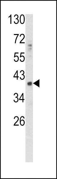
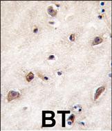
Application
| IHC-P, WB, E |
|---|---|
| Primary Accession | P27539 |
| Reactivity | Human |
| Host | Rabbit |
| Clonality | Polyclonal |
| Isotype | Rabbit IgG |
| Calculated MW | 39475 Da |
| Antigen Region | 30-59 aa |
| Gene ID | 2657 |
|---|---|
| Other Names | Embryonic growth/differentiation factor 1, GDF-1, GDF1 |
| Target/Specificity | This GDF1 antibody is generated from rabbits immunized with a KLH conjugated synthetic peptide between 30-59 amino acids from the N-terminal region of human GDF1. |
| Dilution | IHC-P~~1:10~50 WB~~1:1000 E~~Use at an assay dependent concentration. |
| Format | Purified polyclonal antibody supplied in PBS with 0.09% (W/V) sodium azide. This antibody is prepared by Saturated Ammonium Sulfate (SAS) precipitation followed by dialysis against PBS. |
| Storage | Maintain refrigerated at 2-8°C for up to 2 weeks. For long term storage store at -20°C in small aliquots to prevent freeze-thaw cycles. |
| Precautions | GDF1 Antibody (N-term) is for research use only and not for use in diagnostic or therapeutic procedures. |
| Name | GDF1 |
|---|---|
| Function | May mediate cell differentiation events during embryonic development. |
| Cellular Location | Secreted. |
| Tissue Location | Expressed in the brain. |

Thousands of laboratories across the world have published research that depended on the performance of antibodies from Abcepta to advance their research. Check out links to articles that cite our products in major peer-reviewed journals, organized by research category.
info@abcepta.com, and receive a free "I Love Antibodies" mug.

Provided below are standard protocols that you may find useful for product applications.
Background
GDF1 is a member of the bone morphogenetic protein (BMP) family and the TGF-beta superfamily. This group of proteins is characterized by a polybasic proteolytic processing site which is cleaved to produce a mature protein containing seven conserved cysteine residues. The members of this family are regulators of cell growth and differentiation in both embryonic and adult tissues. Studies in rodents suggest that this protein is involved in the establishment of left-right assymetry in early embryogenesis and in neural development in later embryogenesis. This protein is transcribed from a bicistronic mRNA which also encodes the longevity assurance gene.
References
Ducy, P., et al., Kidney Int. 57(6):2207-2214 (2000). Jiang, J.C., et al., Genome Res. 8(12):1259-1272 (1998). Polymeropoulos, M.H., et al., Nat. Genet. 4(4):381-386 (1993). Lee, S.J., Proc. Natl. Acad. Sci. U.S.A. 88(10):4250-4254 (1991).
If you have used an Abcepta product and would like to share how it has performed, please click on the "Submit Review" button and provide the requested information. Our staff will examine and post your review and contact you if needed.
If you have any additional inquiries please email technical services at tech@abcepta.com.
Ordering Information
Other Products
Shipping Information
