


 Foundational characteristics of cancer include proliferation, angiogenesis, migration, evasion of apoptosis, and cellular immortality. Find key markers for these cellular processes and antibodies to detect them.
 The SUMOplot™ Analysis Program predicts and scores sumoylation sites in your protein. SUMOylation is a post-translational modification involved in various cellular processes, such as nuclear-cytosolic transport, transcriptional regulation, apoptosis, protein stability, response to stress, and progression through the cell cycle.
 The Autophagy Receptor Motif Plotter predicts and scores autophagy receptor binding sites in your protein. Identifying proteins connected to this pathway is critical to understanding the role of autophagy in physiological as well as pathological processes such as development, differentiation, neurodegenerative diseases, stress, infection, and cancer.
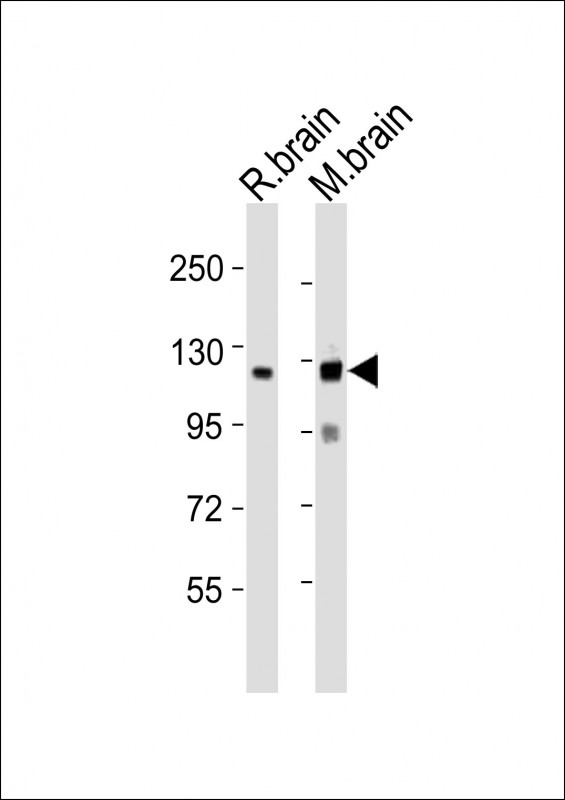
NLGN3 Antibody (C-term)
Purified Rabbit Polyclonal Antibody (Pab)
- SPECIFICATION
- CITATIONS
- PROTOCOLS
- BACKGROUND

Application
| WB, E |
|---|---|
| Primary Accession | Q9NZ94 |
| Reactivity | Mouse, Rat |
| Host | Rabbit |
| Clonality | polyclonal |
| Isotype | Rabbit IgG |
| Calculated MW | 93895 Da |
| Gene ID | 54413 |
|---|---|
| Other Names | Neuroligin-3, Gliotactin homolog, NLGN3, KIAA1480, NL3 |
| Target/Specificity | This NLGN3 antibody is generated from a rabbit immunized with a KLH conjugated synthetic peptide between 651-685 amino acids from the C-terminal region of human NLGN3. |
| Dilution | WB~~1:1000 E~~Use at an assay dependent concentration. |
| Format | Purified polyclonal antibody supplied in PBS with 0.09% (W/V) sodium azide. This antibody is purified through a protein A column, followed by peptide affinity purification. |
| Storage | Maintain refrigerated at 2-8°C for up to 2 weeks. For long term storage store at -20°C in small aliquots to prevent freeze-thaw cycles. |
| Precautions | NLGN3 Antibody (C-term) is for research use only and not for use in diagnostic or therapeutic procedures. |
| Name | NLGN3 |
|---|---|
| Synonyms | KIAA1480, NL3 |
| Function | Cell surface protein involved in cell-cell-interactions via its interactions with neurexin family members. Plays a role in synapse function and synaptic signal transmission, and may mediate its effects by clustering other synaptic proteins. May promote the initial formation of synapses, but is not essential for this. May also play a role in glia-glia or glia-neuron interactions in the developing peripheral nervous system (By similarity). |
| Cellular Location | Cell membrane; Single-pass type I membrane protein. Synapse. Note=Detected at both glutamatergic and GABAergic synapses. |
| Tissue Location | Expressed in the blood vessel walls (at protein level). Detected in throughout the brain and in spinal cord. Detected in brain, and at lower levels in pancreas islet beta cells |

Thousands of laboratories across the world have published research that depended on the performance of antibodies from Abcepta to advance their research. Check out links to articles that cite our products in major peer-reviewed journals, organized by research category.
info@abcepta.com, and receive a free "I Love Antibodies" mug.

Provided below are standard protocols that you may find useful for product applications.
Background
Cell surface protein involved in cell-cell-interactions via its interactions with neurexin family members. Plays a role in synapse function and synaptic signal transmission, and may mediate its effects by clustering other synaptic proteins. May promote the initial formation of synapses, but is not essential for this. May also play a role in glia-glia or glia-neuron interactions in the developing peripheral nervous system (By similarity).
References
Philibert R.A.,et al.Gene 246:303-310(2000).
Rissone A.,et al.Dev. Dyn. 239:688-702(2010).
Ota T.,et al.Nat. Genet. 36:40-45(2004).
Ross M.T.,et al.Nature 434:325-337(2005).
Mural R.J.,et al.Submitted (SEP-2005) to the EMBL/GenBank/DDBJ databases.
If you have used an Abcepta product and would like to share how it has performed, please click on the "Submit Review" button and provide the requested information. Our staff will examine and post your review and contact you if needed.
If you have any additional inquiries please email technical services at tech@abcepta.com.
Ordering Information
Shipping Information
