


 Foundational characteristics of cancer include proliferation, angiogenesis, migration, evasion of apoptosis, and cellular immortality. Find key markers for these cellular processes and antibodies to detect them.
 The SUMOplot™ Analysis Program predicts and scores sumoylation sites in your protein. SUMOylation is a post-translational modification involved in various cellular processes, such as nuclear-cytosolic transport, transcriptional regulation, apoptosis, protein stability, response to stress, and progression through the cell cycle.
 The Autophagy Receptor Motif Plotter predicts and scores autophagy receptor binding sites in your protein. Identifying proteins connected to this pathway is critical to understanding the role of autophagy in physiological as well as pathological processes such as development, differentiation, neurodegenerative diseases, stress, infection, and cancer.
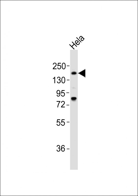
LAMA4 Antibody (C-term)
Purified Rabbit Polyclonal Antibody (Pab)
- SPECIFICATION
- CITATIONS
- PROTOCOLS
- BACKGROUND

Application
| WB, E |
|---|---|
| Primary Accession | Q16363 |
| Reactivity | Human |
| Host | Rabbit |
| Clonality | polyclonal |
| Isotype | Rabbit IgG |
| Calculated MW | 202568 Da |
| Gene ID | 3910 |
|---|---|
| Other Names | Laminin subunit alpha-4, Laminin-14 subunit alpha, Laminin-8 subunit alpha, Laminin-9 subunit alpha, LAMA4 |
| Target/Specificity | This LAMA4 antibody is generated from a rabbit immunized with a KLH conjugated synthetic peptide between 1474-1508 amino acids from the C-terminal region of human LAMA4. |
| Dilution | WB~~1:2000 E~~Use at an assay dependent concentration. |
| Format | Purified polyclonal antibody supplied in PBS with 0.09% (W/V) sodium azide. This antibody is purified through a protein A column, followed by peptide affinity purification. |
| Storage | Maintain refrigerated at 2-8°C for up to 2 weeks. For long term storage store at -20°C in small aliquots to prevent freeze-thaw cycles. |
| Precautions | LAMA4 Antibody (C-term) is for research use only and not for use in diagnostic or therapeutic procedures. |
| Name | LAMA4 |
|---|---|
| Function | Binding to cells via a high affinity receptor, laminin is thought to mediate the attachment, migration and organization of cells into tissues during embryonic development by interacting with other extracellular matrix components. |
| Cellular Location | Secreted, extracellular space, extracellular matrix, basement membrane. Secreted. Note=Major basement membrane component |
| Tissue Location | Detected in placenta (at protein level) (PubMed:32337544). Detected in fibroblasts and urine (at protein level) (PubMed:25326458, PubMed:36213313, PubMed:37453717). In adult, strong expression in heart, lung, ovary small and large intestines, placenta, liver; weak or no expression in skeletal muscle, kidney, pancreas, testis, prostate, brain. High expression in fetal lung and kidney Expression in fetal and newborn tissues is observed in certain mesenchymal cells in tissues such as smooth muscle and dermis |

Thousands of laboratories across the world have published research that depended on the performance of antibodies from Abcepta to advance their research. Check out links to articles that cite our products in major peer-reviewed journals, organized by research category.
info@abcepta.com, and receive a free "I Love Antibodies" mug.

Provided below are standard protocols that you may find useful for product applications.
Background
Binding to cells via a high affinity receptor, laminin is thought to mediate the attachment, migration and organization of cells into tissues during embryonic development by interacting with other extracellular matrix components.
References
Iivanainen A.,et al.FEBS Lett. 365:183-188(1995).
Richards A.J.,et al.Eur. J. Biochem. 238:813-821(1996).
Nakajima D.,et al.Submitted (MAR-2005) to the EMBL/GenBank/DDBJ databases.
Kalnine N.,et al.Submitted (MAY-2003) to the EMBL/GenBank/DDBJ databases.
Mungall A.J.,et al.Nature 425:805-811(2003).
If you have used an Abcepta product and would like to share how it has performed, please click on the "Submit Review" button and provide the requested information. Our staff will examine and post your review and contact you if needed.
If you have any additional inquiries please email technical services at tech@abcepta.com.
Ordering Information
Shipping Information
