


 Foundational characteristics of cancer include proliferation, angiogenesis, migration, evasion of apoptosis, and cellular immortality. Find key markers for these cellular processes and antibodies to detect them.
 The SUMOplot™ Analysis Program predicts and scores sumoylation sites in your protein. SUMOylation is a post-translational modification involved in various cellular processes, such as nuclear-cytosolic transport, transcriptional regulation, apoptosis, protein stability, response to stress, and progression through the cell cycle.
 The Autophagy Receptor Motif Plotter predicts and scores autophagy receptor binding sites in your protein. Identifying proteins connected to this pathway is critical to understanding the role of autophagy in physiological as well as pathological processes such as development, differentiation, neurodegenerative diseases, stress, infection, and cancer.
NRXN1 Antibody (N-term)
Purified Rabbit Polyclonal Antibody (Pab)
- SPECIFICATION
- CITATIONS
- PROTOCOLS
- BACKGROUND

Application
| WB, E |
|---|---|
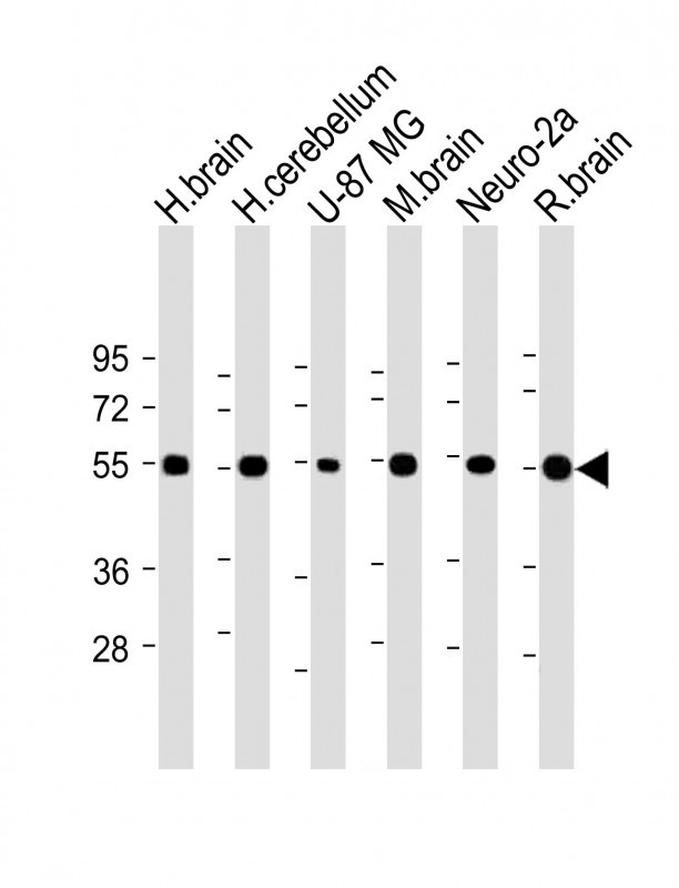
| Primary Accession | P58400 |
| Reactivity | Human, Mouse, Rat |
| Host | Rabbit |
| Clonality | polyclonal |
| Isotype | Rabbit IgG |
| Calculated MW | 50424 Da |
| Antigen Region | 77-110 aa |
| Gene ID | 9378 |
|---|---|
| Other Names | Neurexin-1-beta, Neurexin I-beta, NRXN1 |
| Target/Specificity | This NRXN1 antibody is generated from a rabbit immunized with a KLH conjugated synthetic peptide between 77-110 amino acids from the N-terminal region of human NRXN1. |
| Dilution | WB~~1:2000 E~~Use at an assay dependent concentration. |
| Format | Purified polyclonal antibody supplied in PBS with 0.09% (W/V) sodium azide. This antibody is purified through a protein A column, followed by peptide affinity purification. |
| Storage | Maintain refrigerated at 2-8°C for up to 2 weeks. For long term storage store at -20°C in small aliquots to prevent freeze-thaw cycles. |
| Precautions | NRXN1 Antibody (N-term) is for research use only and not for use in diagnostic or therapeutic procedures. |
| Name | NRXN1 (HGNC:8008) |
|---|---|
| Function | Neuronal cell surface protein involved in cell recognition and cell adhesion by forming intracellular junctions through binding to neuroligins (By similarity). Plays a role in formation of synaptic junctions (By similarity). Functions as part of a trans-synaptic complex by binding to cerebellins and postsynaptic GRID1. This interaction helps regulate the activity of NMDA and AMPA receptors at hippocampal synapses without affecting synapse formation. NRXN1B-CBLN2- GRID1 complex transduce presynaptic signals into postsynaptic NMDAR response (By similarity). |
| Cellular Location | Presynaptic cell membrane {ECO:0000250|UniProtKB:P0DI97}; Single-pass type I membrane protein |

Thousands of laboratories across the world have published research that depended on the performance of antibodies from Abcepta to advance their research. Check out links to articles that cite our products in major peer-reviewed journals, organized by research category.
info@abcepta.com, and receive a free "I Love Antibodies" mug.

Provided below are standard protocols that you may find useful for product applications.
Background
Neuronal cell surface protein that may be involved in cell recognition and cell adhesion by forming intracellular junctions through binding to neuroligins. May play a role in formation or maintenance of synaptic junctions. May mediate intracellular signaling. May play a role in angiogenesis (By similarity).
References
Kleiderlein J.J.,et al.Hum. Genet. 103:666-673(1998).
Hillier L.W.,et al.Nature 434:724-731(2005).
Chen X.,et al.Nat. Struct. Mol. Biol. 15:50-56(2008).
If you have used an Abcepta product and would like to share how it has performed, please click on the "Submit Review" button and provide the requested information. Our staff will examine and post your review and contact you if needed.
If you have any additional inquiries please email technical services at tech@abcepta.com.
Ordering Information
Shipping Information
