


 Foundational characteristics of cancer include proliferation, angiogenesis, migration, evasion of apoptosis, and cellular immortality. Find key markers for these cellular processes and antibodies to detect them.
 The SUMOplot™ Analysis Program predicts and scores sumoylation sites in your protein. SUMOylation is a post-translational modification involved in various cellular processes, such as nuclear-cytosolic transport, transcriptional regulation, apoptosis, protein stability, response to stress, and progression through the cell cycle.
 The Autophagy Receptor Motif Plotter predicts and scores autophagy receptor binding sites in your protein. Identifying proteins connected to this pathway is critical to understanding the role of autophagy in physiological as well as pathological processes such as development, differentiation, neurodegenerative diseases, stress, infection, and cancer.
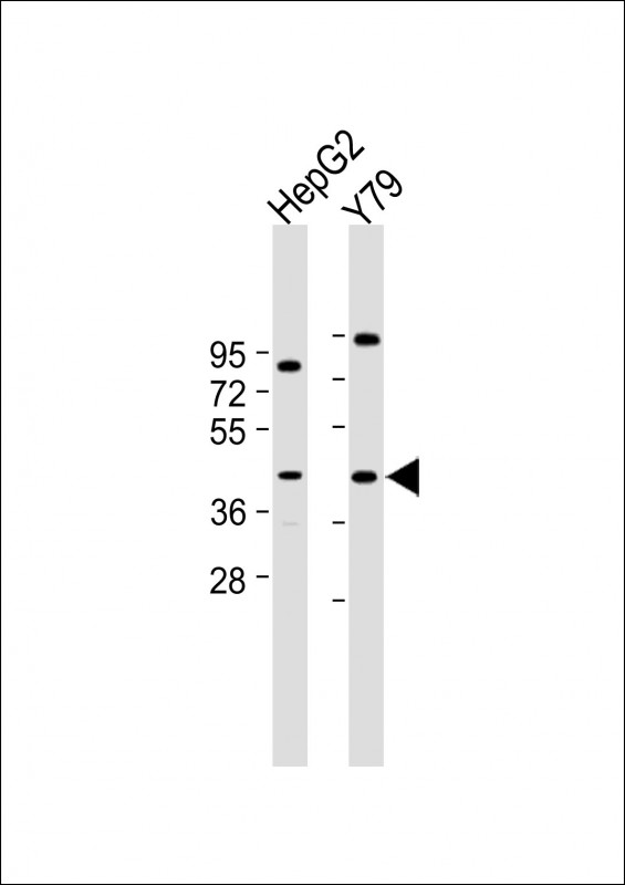
PTGDR Antibody (C-Term)
Purified Rabbit Polyclonal Antibody (Pab)
- SPECIFICATION
- CITATIONS
- PROTOCOLS
- BACKGROUND

Application
| WB, E |
|---|---|
| Primary Accession | Q13258 |
| Reactivity | Human |
| Host | Rabbit |
| Clonality | polyclonal |
| Isotype | Rabbit IgG |
| Calculated MW | 40271 Da |
| Gene ID | 5729 |
|---|---|
| Other Names | Prostaglandin D2 receptor, PGD receptor, PGD2 receptor, Prostanoid DP receptor, PTGDR |
| Target/Specificity | This PTGDR antibody is generated from a rabbit immunized with a KLH conjugated synthetic peptide between 306-348 amino acids from human PTGDR. |
| Dilution | WB~~1:2000 E~~Use at an assay dependent concentration. |
| Format | Purified polyclonal antibody supplied in PBS with 0.09% (W/V) sodium azide. This antibody is purified through a protein A column, followed by peptide affinity purification. |
| Storage | Maintain refrigerated at 2-8°C for up to 2 weeks. For long term storage store at -20°C in small aliquots to prevent freeze-thaw cycles. |
| Precautions | PTGDR Antibody (C-Term) is for research use only and not for use in diagnostic or therapeutic procedures. |
| Name | PTGDR |
|---|---|
| Function | Receptor for prostaglandin D2 (PGD2). The activity of this receptor is mainly mediated by G(s) proteins that stimulate adenylate cyclase, resulting in an elevation of intracellular cAMP. A mobilization of calcium is also observed, but without formation of inositol 1,4,5-trisphosphate (By similarity). Involved in PLA2G3- dependent maturation of mast cells. PLA2G3 is secreted by immature mast cells and acts on nearby fibroblasts upstream to PTDGS to synthesize PGD2, which in turn promotes mast cell maturation and degranulation via PTGDR (By similarity). |
| Cellular Location | Cell membrane; Multi-pass membrane protein |
| Tissue Location | Expressed in retinal choroid, ciliary epithelium, longitudinal and circular ciliary muscles, iris, small intestine and platelet membranes. |

Thousands of laboratories across the world have published research that depended on the performance of antibodies from Abcepta to advance their research. Check out links to articles that cite our products in major peer-reviewed journals, organized by research category.
info@abcepta.com, and receive a free "I Love Antibodies" mug.

Provided below are standard protocols that you may find useful for product applications.
Background
Receptor for prostaglandin D2 (PGD2). The activity of this receptor is mainly mediated by G(s) proteins that stimulate adenylate cyclase, resulting in an elevation of intracellular cAMP. A mobilization of calcium is also observed, but without formation of inositol 1,4,5-trisphosphate (By similarity).
References
Boie Y.,et al.J. Biol. Chem. 270:18910-18916(1995).
Martin A.L.,et al.Submitted (APR-2007) to the EMBL/GenBank/DDBJ databases.
Heilig R.,et al.Nature 421:601-607(2003).
Mural R.J.,et al.Submitted (SEP-2005) to the EMBL/GenBank/DDBJ databases.
Town M.H.,et al.Prostaglandins 25:13-28(1983).
If you have used an Abcepta product and would like to share how it has performed, please click on the "Submit Review" button and provide the requested information. Our staff will examine and post your review and contact you if needed.
If you have any additional inquiries please email technical services at tech@abcepta.com.
Ordering Information
Other Products
Shipping Information
