


 Foundational characteristics of cancer include proliferation, angiogenesis, migration, evasion of apoptosis, and cellular immortality. Find key markers for these cellular processes and antibodies to detect them.
 The SUMOplot™ Analysis Program predicts and scores sumoylation sites in your protein. SUMOylation is a post-translational modification involved in various cellular processes, such as nuclear-cytosolic transport, transcriptional regulation, apoptosis, protein stability, response to stress, and progression through the cell cycle.
 The Autophagy Receptor Motif Plotter predicts and scores autophagy receptor binding sites in your protein. Identifying proteins connected to this pathway is critical to understanding the role of autophagy in physiological as well as pathological processes such as development, differentiation, neurodegenerative diseases, stress, infection, and cancer.
Phospho-nNOS(S1417) Antibody
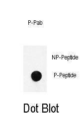
Purified Rabbit Polyclonal Antibody (Pab)
- SPECIFICATION
- CITATIONS
- PROTOCOLS
- BACKGROUND

Application
| WB, DB, E |
|---|---|
| Primary Accession | P29475 |
| Other Accession | P29476, O19132, Q9Z0J4, Q29498 |
| Reactivity | Human |
| Predicted | Mouse, Rabbit, Rat, Sheep |
| Host | Rabbit |
| Clonality | Polyclonal |
| Isotype | Rabbit IgG |
| Calculated MW | 160970 Da |
| Gene ID | 4842 |
|---|---|
| Other Names | Nitric oxide synthase, brain, Constitutive NOS, NC-NOS, NOS type I, Neuronal NOS, N-NOS, nNOS, Peptidyl-cysteine S-nitrosylase NOS1, bNOS, NOS1 |
| Target/Specificity | This nNOS Antibody is generated from rabbits immunized with a KLH conjugated synthetic phosphopeptide corresponding to amino acid residues surrounding S1417 of human nNOS. |
| Dilution | WB~~1:1000 DB~~1:500 E~~Use at an assay dependent concentration. |
| Format | Purified polyclonal antibody supplied in PBS with 0.09% (W/V) sodium azide. This antibody is purified through a protein A column, followed by peptide affinity purification. |
| Storage | Maintain refrigerated at 2-8°C for up to 2 weeks. For long term storage store at -20°C in small aliquots to prevent freeze-thaw cycles. |
| Precautions | Phospho-nNOS(S1417) Antibody is for research use only and not for use in diagnostic or therapeutic procedures. |
| Name | NOS1 (HGNC:7872) |
|---|---|
| Function | Produces nitric oxide (NO) which is a messenger molecule with diverse functions throughout the body. In the brain and peripheral nervous system, NO displays many properties of a neurotransmitter. Probably has nitrosylase activity and mediates cysteine S-nitrosylation of cytoplasmic target proteins such SRR. |
| Cellular Location | Cell membrane, sarcolemma {ECO:0000250|UniProtKB:Q9Z0J4}; Peripheral membrane protein. Cell projection, dendritic spine {ECO:0000250|UniProtKB:P29476}. Note=In skeletal muscle, it is localized beneath the sarcolemma of fast-twitch muscle fiber by associating with the dystrophin glycoprotein complex (By similarity) In neurons, enriched in dendritic spines (By similarity) {ECO:0000250|UniProtKB:P29476, ECO:0000250|UniProtKB:Q9Z0J4} |
| Tissue Location | Isoform 1 is ubiquitously expressed: detected in skeletal muscle and brain, also in testis, lung and kidney, and at low levels in heart, adrenal gland and retina. Not detected in the platelets. Isoform 3 is expressed only in testis. Isoform 4 is detected in testis, skeletal muscle, lung, and kidney, at low levels in the brain, but not in the heart and adrenal gland |

Thousands of laboratories across the world have published research that depended on the performance of antibodies from Abcepta to advance their research. Check out links to articles that cite our products in major peer-reviewed journals, organized by research category.
info@abcepta.com, and receive a free "I Love Antibodies" mug.

Provided below are standard protocols that you may find useful for product applications.
Background
Three isoforms of nitric oxide synthase (NOS) have been identified. All are homodimers with subunits of 130-160 kDa. All have binding sites for NADPH, FAD, and FMN near the carboxyl terminus (the reductase domain), and binding sites for tetrahydrobiopterin (BH4) and heme near the amino terminus (the oxygenase domain). The reductase and oxygenase domains are linked by a calmodulin (CaM) binding site. Occupation of this site facilitates electron transfer from the cofactors in the reductase domain to heme during nitric oxide production. NOS catalyzes the conversion of arginine to citrulline and nitric oxide (NO). Neuronal nitric oxide synthase (nNOS, bNOS, cNOS, Type I) is associated with the post-synaptic density protein (PSD-95) in the neuronal membrane. In response to increased intracellular Ca2+, nNOS interacts with CaM. The Ca2+ CaM complex, in combination with BH4, binds to nNOS and induces its translocation from the plasma membrane to the cytoplasm. The dephosphorylation of nNOS by calcineurin initiates the production NO. NO activates guanylyl cyclase (GC) and activates the various cGMP regulated signaling pathways. nNOS is in activated by phosphorylation by protein kinase A (PKA) or protein kinase C (PKC).
References
Laas,K., et.al., Psychopharmacology (Berl.) 209 (3), 255-261 (2010)
Darrah,R., et.al., Physiol. Genomics 41 (1), 71-77 (2010)
If you have used an Abcepta product and would like to share how it has performed, please click on the "Submit Review" button and provide the requested information. Our staff will examine and post your review and contact you if needed.
If you have any additional inquiries please email technical services at tech@abcepta.com.
Ordering Information
Other Products
Shipping Information
