


 Foundational characteristics of cancer include proliferation, angiogenesis, migration, evasion of apoptosis, and cellular immortality. Find key markers for these cellular processes and antibodies to detect them.
 The SUMOplot™ Analysis Program predicts and scores sumoylation sites in your protein. SUMOylation is a post-translational modification involved in various cellular processes, such as nuclear-cytosolic transport, transcriptional regulation, apoptosis, protein stability, response to stress, and progression through the cell cycle.
 The Autophagy Receptor Motif Plotter predicts and scores autophagy receptor binding sites in your protein. Identifying proteins connected to this pathway is critical to understanding the role of autophagy in physiological as well as pathological processes such as development, differentiation, neurodegenerative diseases, stress, infection, and cancer.
ATL3 Antibody (Center)
Affinity Purified Rabbit Polyclonal Antibody (Pab)
- SPECIFICATION
- CITATIONS
- PROTOCOLS
- BACKGROUND

Application
| WB, E |
|---|---|
| Primary Accession | Q6DD88 |
| Other Accession | Q0ZHH6 |
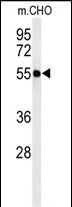
| Reactivity | Human, Hamster, Mouse |
| Predicted | Rat |
| Host | Rabbit |
| Clonality | Polyclonal |
| Isotype | Rabbit IgG |
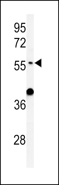
| Calculated MW | 60542 Da |
| Antigen Region | 217-245 aa |
| Gene ID | 25923 |
|---|---|
| Other Names | Atlastin-3, 365-, ATL3 |
| Target/Specificity | This ATL3 antibody is generated from rabbits immunized with a KLH conjugated synthetic peptide between 217-245 amino acids from the Central region of human ATL3. |
| Dilution | WB~~1:1000 E~~Use at an assay dependent concentration. |
| Format | Purified polyclonal antibody supplied in PBS with 0.09% (W/V) sodium azide. This antibody is purified through a protein A column, followed by peptide affinity purification. |
| Storage | Maintain refrigerated at 2-8°C for up to 2 weeks. For long term storage store at -20°C in small aliquots to prevent freeze-thaw cycles. |
| Precautions | ATL3 Antibody (Center) is for research use only and not for use in diagnostic or therapeutic procedures. |
| Name | ATL3 (HGNC:24526) |
|---|---|
| Function | Atlastin-3 (ATL3) is a membrane-anchored GTPase that mediates the GTP-dependent fusion of endoplasmic reticulum (ER) membranes, maintaining the continuous ER network. It facilitates the formation of three-way junctions where ER tubules intersect (PubMed:18270207, PubMed:19665976, PubMed:24459106, PubMed:27619977, PubMed:37102997). Two atlastin-3 on neighboring ER tubules bind GTP and form loose homodimers through the GB1/RHD3-type G domains and 3HB regions. Upon GTP hydrolysis, the 3HB regions tighten, pulling the membranes together to drive their fusion. After fusion, the homodimer disassembles upon release of inorganic phosphate (Pi). Subsequently, GDP dissociates, resetting the monomers to a conformation ready for a new fusion cycle (By similarity). |
| Cellular Location | Endoplasmic reticulum membrane; Multi-pass membrane protein. Note=Localizes to endoplasmic reticulum tubules and accumulates in punctuate structures corresponding to 3-way junctions, which represent crossing-points at which the tubules build a polygonal network. |
| Tissue Location | Expressed in the central nervous system and in dorsal root ganglia neurons. Expressed in peripheral tissues (at protein level). |

Thousands of laboratories across the world have published research that depended on the performance of antibodies from Abcepta to advance their research. Check out links to articles that cite our products in major peer-reviewed journals, organized by research category.
info@abcepta.com, and receive a free "I Love Antibodies" mug.

Provided below are standard protocols that you may find useful for product applications.
Background
ATL3 is GTPase tethering membranes through formation of trans-homooligomer and mediating homotypic fusion of endoplasmic reticulum membranes. ATL3 is function in endoplasmic reticulum tubular network biogenesis.
References
Hu, J., et al. Cell 138(3):549-561(2009)
Rismanchi, N., et al. Hum. Mol. Genet. 17(11):1591-1604(2008)
If you have used an Abcepta product and would like to share how it has performed, please click on the "Submit Review" button and provide the requested information. Our staff will examine and post your review and contact you if needed.
If you have any additional inquiries please email technical services at tech@abcepta.com.
Ordering Information
Other Products
Shipping Information
