


 Foundational characteristics of cancer include proliferation, angiogenesis, migration, evasion of apoptosis, and cellular immortality. Find key markers for these cellular processes and antibodies to detect them.
 The SUMOplot™ Analysis Program predicts and scores sumoylation sites in your protein. SUMOylation is a post-translational modification involved in various cellular processes, such as nuclear-cytosolic transport, transcriptional regulation, apoptosis, protein stability, response to stress, and progression through the cell cycle.
 The Autophagy Receptor Motif Plotter predicts and scores autophagy receptor binding sites in your protein. Identifying proteins connected to this pathway is critical to understanding the role of autophagy in physiological as well as pathological processes such as development, differentiation, neurodegenerative diseases, stress, infection, and cancer.
BHLHB5 Antibody (Center)
Affinity Purified Rabbit Polyclonal Antibody (Pab)
- SPECIFICATION
- CITATIONS
- PROTOCOLS
- BACKGROUND

Application
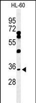
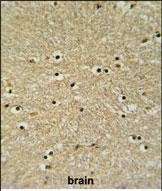
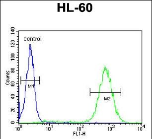
| FC, IHC-P, WB, E |
|---|---|
| Primary Accession | Q8NFJ8 |
| Other Accession | Q8C6A8, Q71T09 |
| Reactivity | Human |
| Predicted | Chicken, Mouse |
| Host | Rabbit |
| Clonality | Polyclonal |
| Isotype | Rabbit IgG |
| Calculated MW | 36997 Da |
| Antigen Region | 236-264 aa |
| Gene ID | 27319 |
|---|---|
| Other Names | Class E basic helix-loop-helix protein 22, bHLHe22, Class B basic helix-loop-helix protein 5, bHLHb5, Trinucleotide repeat-containing gene 20 protein, BHLHE22, BHLHB5, TNRC20 |
| Target/Specificity | This BHLHB5 antibody is generated from rabbits immunized with a KLH conjugated synthetic peptide between 236-264 amino acids from the Central region of human BHLHB5. |
| Dilution | FC~~1:10~50 IHC-P~~1:50~100 WB~~1:1000 E~~Use at an assay dependent concentration. |
| Format | Purified polyclonal antibody supplied in PBS with 0.09% (W/V) sodium azide. This antibody is purified through a protein A column, followed by peptide affinity purification. |
| Storage | Maintain refrigerated at 2-8°C for up to 2 weeks. For long term storage store at -20°C in small aliquots to prevent freeze-thaw cycles. |
| Precautions | BHLHB5 Antibody (Center) is for research use only and not for use in diagnostic or therapeutic procedures. |
| Name | BHLHE22 |
|---|---|
| Synonyms | BHLHB5, TNRC20 |
| Function | Inhibits DNA binding of TCF3/E47 homodimers and TCF3 (E47)/NEUROD1 heterodimers and acts as a strong repressor of Neurod1 and Myod-responsive genes, probably by heterodimerization with class a basic helix-loop-helix factors. Despite the presence of an intact basic domain, does not bind to DNA (By similarity). In the brain, may function as an area-specific transcription factor that regulates the postmitotic acquisition of area identities and elucidate the genetic hierarchy between progenitors and postmitotic neurons driving neocortical arealization. May be required for the survival of a specific population of inhibitory neurons in the superficial laminae of the spinal cord dorsal horn that may regulate pruritis. Seems to play a crucial role in the retinogenesis, in the specification of amacrine and bipolar subtypes. Forms with PRDM8 a transcriptional repressor complex controlling genes involved in neural development and neuronal differentiation. |
| Cellular Location | Nucleus. |
| Tissue Location | Brain-specific, with the highest expression in the cerebellum. |

Thousands of laboratories across the world have published research that depended on the performance of antibodies from Abcepta to advance their research. Check out links to articles that cite our products in major peer-reviewed journals, organized by research category.
info@abcepta.com, and receive a free "I Love Antibodies" mug.

Provided below are standard protocols that you may find useful for product applications.
Background
BHLHB5 inhibits DNA binding of TCF3/E47 homodimers and TCF3 (E47)/NEUROD1 heterodimers and acts as a strong repressor of Neurod1 and Myod-responsive genes, probably by heterodimerization with class a basic helix-loop-helix factors. BHLHB5 despite the presence of an intact basic domain, does not bind to DNA.
References
Stevens, J.D., et al. Differentiation 76(9):1006-1022(2008)
McLellan, A.S., et al. Mech. Dev. 119 SUPPL 1, S285-S291 (2002)
If you have used an Abcepta product and would like to share how it has performed, please click on the "Submit Review" button and provide the requested information. Our staff will examine and post your review and contact you if needed.
If you have any additional inquiries please email technical services at tech@abcepta.com.
Ordering Information
Other Products
Shipping Information
