


 Foundational characteristics of cancer include proliferation, angiogenesis, migration, evasion of apoptosis, and cellular immortality. Find key markers for these cellular processes and antibodies to detect them.
 The SUMOplot™ Analysis Program predicts and scores sumoylation sites in your protein. SUMOylation is a post-translational modification involved in various cellular processes, such as nuclear-cytosolic transport, transcriptional regulation, apoptosis, protein stability, response to stress, and progression through the cell cycle.
 The Autophagy Receptor Motif Plotter predicts and scores autophagy receptor binding sites in your protein. Identifying proteins connected to this pathway is critical to understanding the role of autophagy in physiological as well as pathological processes such as development, differentiation, neurodegenerative diseases, stress, infection, and cancer.
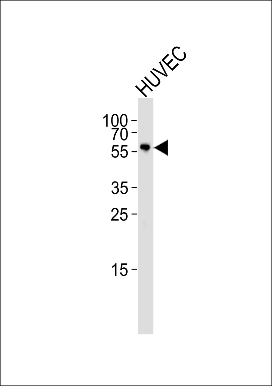
CLK2 Antibody
Purified Rabbit Polyclonal Antibody (Pab)
- SPECIFICATION
- CITATIONS
- PROTOCOLS
- BACKGROUND

Application
| WB |
|---|---|
| Primary Accession | P49760 |
| Reactivity | Human, Mouse, Rat |
| Host | Rabbit |
| Clonality | Polyclonal |
| Calculated MW | 60 18 KDa |
| Antigen Region | 4-35 aa |
| Gene ID | 1196 |
|---|---|
| Other Names | Dual specificity protein kinase CLK2, CDC-like kinase 2, CLK2 |
| Dilution | WB~~1:1000 |
| Format | Rabbit IgG in phosphate buffered saline (without Mg2+ and Ca2+), pH 7.4, 150mM NaCl, 0.09% (W/V) sodium azide and 50% glycerol. |
| Storage Conditions | -20℃ |
| Name | CLK2 |
|---|---|
| Function | Dual specificity kinase acting on both serine/threonine and tyrosine-containing substrates. Phosphorylates serine- and arginine- rich (SR) proteins of the spliceosomal complex. May be a constituent of a network of regulatory mechanisms that enable SR proteins to control RNA splicing and can cause redistribution of SR proteins from speckles to a diffuse nucleoplasmic distribution. Acts as a suppressor of hepatic gluconeogenesis and glucose output by repressing PPARGC1A transcriptional activity on gluconeogenic genes via its phosphorylation. Phosphorylates PPP2R5B thereby stimulating the assembly of PP2A phosphatase with the PPP2R5B-AKT1 complex leading to dephosphorylation of AKT1. Phosphorylates: PTPN1, SRSF1 and SRSF3. Regulates the alternative splicing of tissue factor (F3) pre-mRNA in endothelial cells. Phosphorylates PAGE4 at several serine and threonine residues and this phosphorylation attenuates the ability of PAGE4 to potentiate the transcriptional activator activity of JUN (PubMed:28289210). |
| Cellular Location | Nucleus. [Isoform 2]: Nucleus speckle. Note=Co-localizes with serine- and arginine-rich (SR) proteins in the nuclear speckles |
| Tissue Location | Endothelial cells (PubMed:19168442). Expressed in androgen-dependent prostate cancer cells (PubMed:28289210) |

Thousands of laboratories across the world have published research that depended on the performance of antibodies from Abcepta to advance their research. Check out links to articles that cite our products in major peer-reviewed journals, organized by research category.
info@abcepta.com, and receive a free "I Love Antibodies" mug.

Provided below are standard protocols that you may find useful for product applications.
Background
Dual specificity kinase acting on both serine/threonine and tyrosine-containing substrates. Phosphorylates serine- and arginine-rich (SR) proteins of the spliceosomal complex. May be a constituent of a network of regulatory mechanisms that enable SR proteins to control RNA splicing and can cause redistribution of SR proteins from speckles to a diffuse nucleoplasmic distribution. Acts as a suppressor of hepatic gluconeogenesis and glucose output by repressing PPARGC1A transcriptional activity on gluconeogenic genes via its phosphorylation. Phosphorylates PPP2R5B thereby stimulating the assembly of PP2A phosphatase with the PPP2R5B-AKT1 complex leading to dephosphorylation of AKT1. Phosphorylates: PTPN1, SRSF1 and SRSF3. Regulates the alternative splicing of tissue factor (F3) pre-mRNA in endothelial cells.
References
Hanes J.J.,et al.J. Mol. Biol. 244:665-672(1994).
Winfield S.L.,et al.Genome Res. 7:1020-1026(1997).
Gregory S.G.,et al.Nature 441:315-321(2006).
Lee K.,et al.J. Biol. Chem. 271:27299-27303(1996).
Duncan P.I.,et al.Exp. Cell Res. 241:300-308(1998).
If you have used an Abcepta product and would like to share how it has performed, please click on the "Submit Review" button and provide the requested information. Our staff will examine and post your review and contact you if needed.
If you have any additional inquiries please email technical services at tech@abcepta.com.
Ordering Information
Other Products
Shipping Information
