


 Foundational characteristics of cancer include proliferation, angiogenesis, migration, evasion of apoptosis, and cellular immortality. Find key markers for these cellular processes and antibodies to detect them.
 The SUMOplot™ Analysis Program predicts and scores sumoylation sites in your protein. SUMOylation is a post-translational modification involved in various cellular processes, such as nuclear-cytosolic transport, transcriptional regulation, apoptosis, protein stability, response to stress, and progression through the cell cycle.
 The Autophagy Receptor Motif Plotter predicts and scores autophagy receptor binding sites in your protein. Identifying proteins connected to this pathway is critical to understanding the role of autophagy in physiological as well as pathological processes such as development, differentiation, neurodegenerative diseases, stress, infection, and cancer.
TESC Antibody (C-term)
Affinity Purified Rabbit Polyclonal Antibody (Pab)
- SPECIFICATION
- CITATIONS
- PROTOCOLS
- BACKGROUND

Application
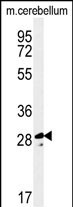
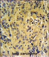
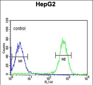
| FC, IHC-P, WB, E |
|---|---|
| Primary Accession | Q96BS2 |
| Reactivity | Human, Mouse |
| Host | Rabbit |
| Clonality | Polyclonal |
| Isotype | Rabbit IgG |
| Calculated MW | 24750 Da |
| Antigen Region | 88-116 aa |
| Gene ID | 54997 |
|---|---|
| Other Names | Calcineurin B homologous protein 3, Tescalcin, TSC, TESC, CHP3 |
| Target/Specificity | This TESC antibody is generated from rabbits immunized with a KLH conjugated synthetic peptide between 88-116 amino acids from the C-terminal region of human TESC. |
| Dilution | FC~~1:10~50 IHC-P~~1:50~100 WB~~1:1000 E~~Use at an assay dependent concentration. |
| Format | Purified polyclonal antibody supplied in PBS with 0.09% (W/V) sodium azide. This antibody is purified through a protein A column, followed by peptide affinity purification. |
| Storage | Maintain refrigerated at 2-8°C for up to 2 weeks. For long term storage store at -20°C in small aliquots to prevent freeze-thaw cycles. |
| Precautions | TESC Antibody (C-term) is for research use only and not for use in diagnostic or therapeutic procedures. |
| Name | TESC |
|---|---|
| Synonyms | CHP3 |
| Function | Functions as an integral cofactor in cell pH regulation by controlling plasma membrane-type Na(+)/H(+) exchange activity. Promotes the maturation, transport, cell surface stability and exchange activity of SLC9A1/NHE1 at the plasma membrane. Promotes the induction of hematopoietic stem cell differentiation toward megakaryocytic lineage. Essential for the coupling of ERK cascade activation with the expression of ETS family genes in megakaryocytic differentiation. Also involved in granulocytic differentiation in a ERK-dependent manner. Inhibits the phosphatase activity of calcineurin. |
| Cellular Location | Nucleus. Cytoplasm. Membrane; Lipid-anchor. Cell membrane. Cell projection, lamellipodium. Cell projection, ruffle membrane {ECO:0000250|UniProtKB:Q9JKL5} Note=Colocalizes with SLC9A1 at the plasma membrane |
| Tissue Location | Expressed in mature megakaryocytes and polymorphonuclear granulocytes (at protein level). Abundantly expressed in heart. Also expressed at a lower level in adult testis and salivary gland, and in the placenta. |

Thousands of laboratories across the world have published research that depended on the performance of antibodies from Abcepta to advance their research. Check out links to articles that cite our products in major peer-reviewed journals, organized by research category.
info@abcepta.com, and receive a free "I Love Antibodies" mug.

Provided below are standard protocols that you may find useful for product applications.
Background
Essential for the coupling of ERK cascade activation with the expression of ETS family genes in megakaryocytic differentiation.
References
Bao, Y., et al. Gene Expr. Patterns 9(5):273-281(2009)
Zaun, H.C., et al. J. Biol. Chem. 283(18):12456-12467(2008)
Levay, K., et al. J. Clin. Invest. 117(9):2672-2683(2007)
If you have used an Abcepta product and would like to share how it has performed, please click on the "Submit Review" button and provide the requested information. Our staff will examine and post your review and contact you if needed.
If you have any additional inquiries please email technical services at tech@abcepta.com.
Ordering Information
Other Products
Shipping Information
