


 Foundational characteristics of cancer include proliferation, angiogenesis, migration, evasion of apoptosis, and cellular immortality. Find key markers for these cellular processes and antibodies to detect them.
 The SUMOplot™ Analysis Program predicts and scores sumoylation sites in your protein. SUMOylation is a post-translational modification involved in various cellular processes, such as nuclear-cytosolic transport, transcriptional regulation, apoptosis, protein stability, response to stress, and progression through the cell cycle.
 The Autophagy Receptor Motif Plotter predicts and scores autophagy receptor binding sites in your protein. Identifying proteins connected to this pathway is critical to understanding the role of autophagy in physiological as well as pathological processes such as development, differentiation, neurodegenerative diseases, stress, infection, and cancer.
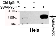
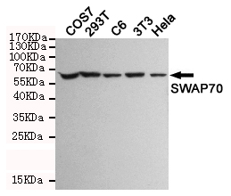
SWAP70 Antibody
Purified Mouse Monoclonal Antibody (Mab)
- SPECIFICATION
- CITATIONS
- PROTOCOLS
- BACKGROUND

Application
| WB, IP |
|---|---|
| Primary Accession | Q9UH65 |
| Reactivity | Human, Mouse |
| Host | Mouse |
| Clonality | Monoclonal |
| Isotype | IgG2b |
| Calculated MW | 70 KDa |
| Gene ID | 23075 |
|---|---|
| Other Names | KIAA0640;70kDa;AV235546;FLJ39540;HSPC321;SWA 70 protein;SWAP 70;SWAP switching B-cell complex 70kDa subunit;SWAP-70;SWAP70;Switch associated protein 70;Switch-associated protein 70;SWP70_HUMAN. |
| Dilution | WB~~1:1000 IP~~1:500 |
| Format | Liquid in PBS containing 50% glycerol, 0.5% BSA and 0.02% sodium azide, pH 7.3. |
| Storage | Store at 4°C short term. Aliquot and store at -20°C long term. Avoid freeze/thaw cycles. |
| Name | SWAP70 |
|---|---|
| Synonyms | KIAA0640 |
| Function | Phosphatidylinositol 3,4,5-trisphosphate-dependent guanine nucleotide exchange factor (GEF) which, independently of RAS, transduces signals from tyrosine kinase receptors to RAC. It also mediates signaling of membrane ruffling. Regulates the actin cytoskeleton as an effector or adapter protein in response to agonist stimulated phosphatidylinositol (3,4)-bisphosphate production and cell protrusion (By similarity). |
| Cellular Location | Cytoplasm. Cell membrane. Nucleus. Cell projection, lamellipodium. Cytoplasm, cytoskeleton. Note=In resting B-cells it is localized mainly in the cytoplasm and upon cell activation it is recruited to the plasma membrane and then translocates to the nucleus (PubMed:10681448) In activated, class-switching B-cells it is associated with membrane IgG but not IgM (PubMed:10681448). Localized to loose actin filament arrays located behind actively extending lamellipodia (PubMed:12925760). |
| Tissue Location | Expressed only in mature B-cells including those associated with mucosa-associated tissue and bronchus-associated tissue (PubMed:10681448). Widely expressed. Abundant in spleen, and fairly abundant in kidney, lung and liver. Also found in monocytes and macrophages (PubMed:12925760). |

Thousands of laboratories across the world have published research that depended on the performance of antibodies from Abcepta to advance their research. Check out links to articles that cite our products in major peer-reviewed journals, organized by research category.
info@abcepta.com, and receive a free "I Love Antibodies" mug.

Provided below are standard protocols that you may find useful for product applications.
Background
Phosphatidylinositol 3,4,5-trisphosphate-dependent guanine nucleotide exchange factor (GEF) which, independently of RAS, transduces signals from tyrosine kinase receptors to RAC. It also mediates signaling of membrane ruffling. Regulates the actin cytoskeleton as an effector or adapter protein in response to agonist stimulated phosphatidylinositol (3,4)-bisphosphate production and cell protrusion (By similarity).
References
Masat L.,et al.Proc. Natl. Acad. Sci. U.S.A. 97:2180-2184(2000).
Rapalus L.,et al.Clin. Immunol. 101:270-275(2001).
Monz D.W.,et al.Submitted (MAR-1999) to the EMBL/GenBank/DDBJ databases.
Ishikawa K.,et al.DNA Res. 5:169-176(1998).
Mural R.J.,et al.Submitted (SEP-2005) to the EMBL/GenBank/DDBJ databases.
If you have used an Abcepta product and would like to share how it has performed, please click on the "Submit Review" button and provide the requested information. Our staff will examine and post your review and contact you if needed.
If you have any additional inquiries please email technical services at tech@abcepta.com.
Ordering Information
Other Products
Shipping Information
