


 Foundational characteristics of cancer include proliferation, angiogenesis, migration, evasion of apoptosis, and cellular immortality. Find key markers for these cellular processes and antibodies to detect them.
 The SUMOplot™ Analysis Program predicts and scores sumoylation sites in your protein. SUMOylation is a post-translational modification involved in various cellular processes, such as nuclear-cytosolic transport, transcriptional regulation, apoptosis, protein stability, response to stress, and progression through the cell cycle.
 The Autophagy Receptor Motif Plotter predicts and scores autophagy receptor binding sites in your protein. Identifying proteins connected to this pathway is critical to understanding the role of autophagy in physiological as well as pathological processes such as development, differentiation, neurodegenerative diseases, stress, infection, and cancer.
OLIG2 Antibody
Purified Mouse Monoclonal Antibody (Mab)
- SPECIFICATION
- CITATIONS
- PROTOCOLS
- BACKGROUND

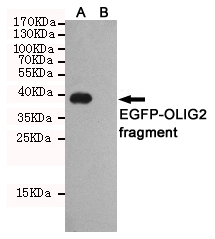
Application
| WB |
|---|---|
| Primary Accession | Q13516 |
| Reactivity | Transfected |
| Host | Mouse |
| Clonality | Monoclonal |
| Isotype | IgG1 |
| Calculated MW | 32 KDa |
| Gene ID | 10215 |
|---|---|
| Other Names | Basic domain helix loop helix protein class B 1; Basic helix loop helix protein class B 1; BHLHB; bHLHB1; bHLHe19; Class B basic helix loop helix protein 1; Class B basic helix-loop-helix protein 1; class E basic helix loop helix protein 19; Class E basic helix-loop-helix protein 19; Human protein kinase C binding protein RACK17; Olig2; OLIG2_HUMAN; Oligo2; Oligodendrocyte lineage transcription factor 2; Oligodendrocyte specific bHLH transcription factor 2; Oligodendrocyte transcription factor 2; OTTHUMP00000067569; OTTHUMP00000067570; PRKCBP2; Protein kinase C binding protein 2; Protein kinase C binding protein RACK17; Protein kinase C-binding protein 2; Protein kinase C-binding protein RACK17; RACK17. |
| Dilution | WB~~1:1000 |
| Format | Liquid in PBS containing 50% glycerol, 0.5% BSA and 0.02% sodium azide, pH 7.3. |
| Storage | Store at 4°C short term. Aliquot and store at -20°C long term. Avoid freeze/thaw cycles. |
| Name | OLIG2 |
|---|---|
| Synonyms | BHLHB1, BHLHE19, PRKCBP2, RACK17 |
| Function | Required for oligodendrocyte and motor neuron specification in the spinal cord, as well as for the development of somatic motor neurons in the hindbrain. Functions together with ZNF488 to promote oligodendrocyte differentiation. Cooperates with OLIG1 to establish the pMN domain of the embryonic neural tube. Antagonist of V2 interneuron and of NKX2-2-induced V3 interneuron development. |
| Cellular Location | Nucleus {ECO:0000255|PROSITE-ProRule:PRU00981}. Cytoplasm. Note=The NLS contained in the bHLH domain could be masked in the native form and translocation to the nucleus could be mediated by interaction either with class E bHLH partner protein or with NKX2-2. |
| Tissue Location | Expressed in the brain, in oligodendrocytes. Strongly expressed in oligodendrogliomas, while expression is weak to moderate in astrocytomas. Expression in glioblastomas highly variable |

Thousands of laboratories across the world have published research that depended on the performance of antibodies from Abcepta to advance their research. Check out links to articles that cite our products in major peer-reviewed journals, organized by research category.
info@abcepta.com, and receive a free "I Love Antibodies" mug.

Provided below are standard protocols that you may find useful for product applications.
Background
Required for oligodendrocyte and motor neuron specification in the spinal cord, as well as for the development of somatic motor neurons in the hindbrain. Cooperates with OLIG1 to establish the pMN domain of the embryonic neural tube. Antagonist of V2 interneuron and of NKX2-2-induced V3 interneuron development (By similarity).
References
Kuroda S.,et al.Submitted (FEB-1996) to the EMBL/GenBank/DDBJ databases.
Wang J.,et al.Proc. Natl. Acad. Sci. U.S.A. 97:3497-3502(2000).
Ota T.,et al.Nat. Genet. 36:40-45(2004).
Marie Y.,et al.Lancet 358:298-300(2001).
Lu Q.R.,et al.Proc. Natl. Acad. Sci. U.S.A. 98:10851-10856(2001).
If you have used an Abcepta product and would like to share how it has performed, please click on the "Submit Review" button and provide the requested information. Our staff will examine and post your review and contact you if needed.
If you have any additional inquiries please email technical services at tech@abcepta.com.

Ordering Information
Other Products
Shipping Information
