


 Foundational characteristics of cancer include proliferation, angiogenesis, migration, evasion of apoptosis, and cellular immortality. Find key markers for these cellular processes and antibodies to detect them.
 The SUMOplot™ Analysis Program predicts and scores sumoylation sites in your protein. SUMOylation is a post-translational modification involved in various cellular processes, such as nuclear-cytosolic transport, transcriptional regulation, apoptosis, protein stability, response to stress, and progression through the cell cycle.
 The Autophagy Receptor Motif Plotter predicts and scores autophagy receptor binding sites in your protein. Identifying proteins connected to this pathway is critical to understanding the role of autophagy in physiological as well as pathological processes such as development, differentiation, neurodegenerative diseases, stress, infection, and cancer.
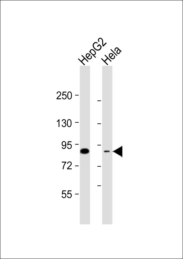
CD248 Antibody
Purified Rabbit Polyclonal Antibody (Pab)
- SPECIFICATION
- CITATIONS
- PROTOCOLS
- BACKGROUND

Application
| WB |
|---|---|
| Primary Accession | Q9HCU0 |
| Reactivity | Human |
| Host | Rabbit |
| Clonality | Polyclonal |
| Calculated MW | 81 KDa |
| Antigen Region | 352-391aa |
| Gene ID | 57124 |
|---|---|
| Other Names | Endosialin, Tumor endothelial marker 1, CD248, CD248, CD164L1, TEM1 |
| Target/Specificity | KLH-conjugated synthetic peptide encompassing a sequence within the center region of human CD248. The exact sequence is proprietary. |
| Dilution | WB~~ 1:1000 |
| Format | Rabbit IgG in phosphate buffered saline , pH 7.4, 150mM NaCl, 0.09% (W/V) sodium azide and 50% glycerol |
| Storage | Store at -20 °C.Stable for 12 months from date of receipt |
| Name | CD248 |
|---|---|
| Synonyms | CD164L1, TEM1 |
| Function | Cell surface glycoprotein involved in various biological processes including angiogenesis, immune response modulation, and tissue remodeling and repair. Participates in pericyte proliferation through positive modulation of the PDGF receptor signaling pathway (PubMed:20484976). Acts as a scaffold for factor X, triggering allosteric changes and the spatial re-alignment of factor X with the TF-factor VIIa complex, thereby enhancing coagulation activation. Modulates the insulin signaling pathway by interacting with insulin receptor/INSR and by diminishing its capacity to be autophosphorylated in response to insulin. Also regulates LPS-induced inflammatory response in macrophages by favoring the production of proinflammatory cytokines. In human, negatively regulates T-cell proliferation compared with stromal cells where it increases proliferation (PubMed:21466550). |
| Cellular Location | Membrane; Single-pass type I membrane protein |
| Tissue Location | Expressed in tumor endothelial cells but absent or barely detectable in normal endothelial cells. Expressed in metastatic lesions of the liver and during angiogenesis of corpus luteum formation and wound healing. Expressed in vascular endothelial cells of malignant tumors but not in normal blood vessels. Expressed in stromal fibroblasts. Strongly expressed in pericytes (PubMed:20484976) Expressed on stromal cells and cells with lymphoid morphology such a T- cells (PubMed:21466550). |

Thousands of laboratories across the world have published research that depended on the performance of antibodies from Abcepta to advance their research. Check out links to articles that cite our products in major peer-reviewed journals, organized by research category.
info@abcepta.com, and receive a free "I Love Antibodies" mug.

Provided below are standard protocols that you may find useful for product applications.
Background
May play a role in tumor angiogenesis.
References
St Croix B.,et al.Science 289:1197-1202(2000).
Christian S.,et al.J. Biol. Chem. 276:7408-7414(2001).
Ota T.,et al.Nat. Genet. 36:40-45(2004).
Rettig W.J.,et al.Proc. Natl. Acad. Sci. U.S.A. 89:10832-10836(1992).
Dolznig H.,et al.Cancer Immun. 5:10-10(2005).
If you have used an Abcepta product and would like to share how it has performed, please click on the "Submit Review" button and provide the requested information. Our staff will examine and post your review and contact you if needed.
If you have any additional inquiries please email technical services at tech@abcepta.com.
Ordering Information
Other Products
Shipping Information
