


 Foundational characteristics of cancer include proliferation, angiogenesis, migration, evasion of apoptosis, and cellular immortality. Find key markers for these cellular processes and antibodies to detect them.
 The SUMOplot™ Analysis Program predicts and scores sumoylation sites in your protein. SUMOylation is a post-translational modification involved in various cellular processes, such as nuclear-cytosolic transport, transcriptional regulation, apoptosis, protein stability, response to stress, and progression through the cell cycle.
 The Autophagy Receptor Motif Plotter predicts and scores autophagy receptor binding sites in your protein. Identifying proteins connected to this pathway is critical to understanding the role of autophagy in physiological as well as pathological processes such as development, differentiation, neurodegenerative diseases, stress, infection, and cancer.
> home > Products > Primary Antibodies > Antibody Collections > Liver positive > Anti-Collagen 18 alpha 1 Antibody
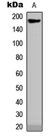
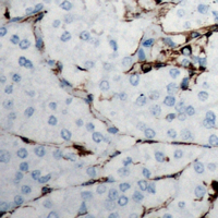
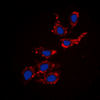
Anti-Collagen 18 alpha 1 Antibody
- SPECIFICATION
- CITATIONS: 1
- PROTOCOLS
- BACKGROUND

Application
| WB, IF, IHC |
|---|---|
| Primary Accession | P39060 |
| Reactivity | Human |
| Host | Rabbit |
| Clonality | Polyclonal |
| Calculated MW | 178188 Da |
| Gene ID | 80781 |
|---|---|
| Other Names | Collagen alpha-1(XVIII) chain |
| Target/Specificity | KLH-conjugated synthetic peptide encompassing a sequence within the center region of human Collagen 18 alpha 1. The exact sequence is proprietary. |
| Dilution | WB~~1/500 - 1/1000 IF~~1/50 - 1/200 IHC~~1:100~500 |
| Format | Liquid in 0.42% Potassium phosphate, 0.87% Sodium chloride, pH 7.3, 30% glycerol, and 0.09% (W/V) sodium azide. |
| Storage | Store at -20 °C.Stable for 12 months from date of receipt |
| Name | COL18A1 (HGNC:2195) |
|---|---|
| Function | Probably plays a major role in determining the retinal structure as well as in the closure of the neural tube. |
| Cellular Location | Secreted, extracellular space, extracellular matrix. Secreted, extracellular space, extracellular matrix, basement membrane {ECO:0000250|UniProtKB:P39061} [Endostatin]: Secreted. Secreted, extracellular space, extracellular matrix, basement membrane |
| Tissue Location | Detected in placenta (at protein level) (PubMed:32337544). Present in multiple organs with highest levels in liver, lung and kidney. |
Research Areas
Citations ( 0 )

Application Protocols
Provided below are standard protocols that you may find useful for product applications.
Background
Rabbit polyclonal antibody to Collagen 18 alpha 1
Abcepta welcomes feedback from its customers.
If you have used an Abcepta product and would like to share how it has performed, please click on the "Submit Review" button and provide the requested information. Our staff will examine and post your review and contact you if needed.
If you have any additional inquiries please email technical services at tech@abcepta.com.
$ 385.00
Cat# AP53893
Ordering Information
United States
AlbaniaAustraliaAustriaBelgiumBosnia & HerzegovinaBrazilBulgariaCanadaCentral AmericaChinaCroatiaCyprusCzech RepublicDenmarkEstoniaFinlandFranceGermanyGreeceHong KongHungaryIcelandIndiaIndonesiaIrelandIsraelItalyJapanLatviaLithuaniaLuxembourgMacedoniaMalaysiaMaltaMexicoNetherlandsNew ZealandNorwayPakistanPolandPortugalRomaniaSerbiaSingaporeSlovakiaSloveniaSouth AfricaSouth KoreaSpainSwedenSwitzerlandTaiwanTurkeyUnited KingdomUnited StatesVietnamWorldwideOthers
USA Headquarters
(888) 735-7227 / (858) 622-0099 or (858) 875-1900
Other Products
Shipping Information
Domestic orders (in stock items)
Shipped out the same day. Orders placed after 1 PM (PST) will ship out the next business day.
International orders
Contact your local distributors
