


 Foundational characteristics of cancer include proliferation, angiogenesis, migration, evasion of apoptosis, and cellular immortality. Find key markers for these cellular processes and antibodies to detect them.
 The SUMOplot™ Analysis Program predicts and scores sumoylation sites in your protein. SUMOylation is a post-translational modification involved in various cellular processes, such as nuclear-cytosolic transport, transcriptional regulation, apoptosis, protein stability, response to stress, and progression through the cell cycle.
 The Autophagy Receptor Motif Plotter predicts and scores autophagy receptor binding sites in your protein. Identifying proteins connected to this pathway is critical to understanding the role of autophagy in physiological as well as pathological processes such as development, differentiation, neurodegenerative diseases, stress, infection, and cancer.
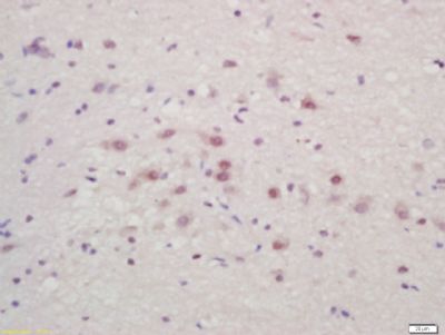
Bassoon Polyclonal Antibody
Purified Rabbit Polyclonal Antibody (Pab)
- SPECIFICATION
- CITATIONS
- PROTOCOLS
- BACKGROUND

Application
| IHC-P, IHC-F, IF |
|---|---|
| Primary Accession | Q9UPA5 |
| Reactivity | Rat |
| Host | Rabbit |
| Clonality | Polyclonal |
| Calculated MW | 432 KDa |
| Physical State | Liquid |
| Immunogen | KLH conjugated synthetic peptide derived from human BSN |
| Epitope Specificity | 2901-3000/3926 |
| Isotype | IgG |
| Purity | affinity purified by Protein A |
| Buffer | 0.01M TBS (pH7.4) with 1% BSA, 0.02% Proclin300 and 50% Glycerol. |
| SUBCELLULAR LOCATION | Cytoplasm. Cell junction, synapse, synaptosome. Cytoplasm, cytoskeleton. Note=Localized to the active zone of presynaptic density. |
| SUBUNIT | Interacts with ERC2/CAST1, RIMS1 and UNC13A. Part of a complex consisting of ERC2, RIMS1 and BSN. |
| Post-translational modifications | Myristoylated. The N-terminal myristoylation is not sufficient for presynaptic localization. |
| Important Note | This product as supplied is intended for research use only, not for use in human, therapeutic or diagnostic applications. |
| Background Descriptions | Neurotransmitters are released from a specific site in the axon terminal called the active zone, which is composed of synaptic vesicles and a meshwork of cytoskeleton underlying the plasma membrane. The protein encoded by this gene is thought to be a scaffolding protein involved in organizing the presynaptic cytoskeleton. The gene is expressed primarily in neurons in the brain. A similar gene product in rodents is concentrated in the active zone of axon terminals and tightly associated with cytoskeletal structures, and is essential for regulating neurotransmitter release from a subset of synapses. [provided by RefSeq, Jul 2008]. |
| Gene ID | 8927 |
|---|---|
| Other Names | Protein bassoon, Zinc finger protein 231, BSN (HGNC:1117) |
| Target/Specificity | Exclusively expressed in brain. |
| Dilution | IHC-P=1:100-500,IHC-F=1:100-500,IF=1:100-500 |
| Storage | Store at -20 ℃ for one year. Avoid repeated freeze/thaw cycles. When reconstituted in sterile pH 7.4 0.01M PBS or diluent of antibody the antibody is stable for at least two weeks at 2-4 ℃. |
| Name | BSN (HGNC:1117) |
|---|---|
| Function | Scaffold protein of the presynaptic cytomatrix at the active zone (CAZ) which is the place in the synapse where neurotransmitter is released (PubMed:12812759). After synthesis, participates in the formation of Golgi-derived membranous organelles termed Piccolo-Bassoon transport vesicles (PTVs) that are transported along axons to sites of nascent synaptic contacts (PubMed:19380881). At the presynaptic active zone, regulates the spatial organization of synaptic vesicle cluster, the protein complexes that execute membrane fusion and compensatory endocytosis (By similarity). Also functions in processes other than assembly such as the regulation of specific presynaptic protein ubiquitination by interacting with SIAH1 or the regulation of presynaptic autophagy by associating with ATG5 (By similarity). Also mediates synapse to nucleus communication leading to reconfiguration of gene expression by associating with the transcriptional corepressor CTBP1 and by subsequently reducing the size of its pool available for nuclear import (By similarity). Inhibits the activity of the proportion of DAO enzyme that localizes to the presynaptic active zone, which may modulate synaptic transmission (By similarity). |
| Cellular Location | Cytoplasm {ECO:0000250|UniProtKB:O88778}. Presynaptic active zone {ECO:0000250|UniProtKB:O88778}. Cytoplasm, cytoskeleton {ECO:0000250|UniProtKB:O88778}. Cytoplasmic vesicle, secretory vesicle, synaptic vesicle membrane {ECO:0000250|UniProtKB:O88778}; Peripheral membrane protein {ECO:0000250|UniProtKB:O88778}. Note=In retina, is localized in the outer plexiform layer at ribbon synapses formed by rods and cones but was absent from basal synaptic contacts formed by cones. In the retinal inner plexiform layer localized to conventional inhibitory GABAergic synapses, made by amacrine cells, but absent from the bipolar cell ribbon synapses. {ECO:0000250|UniProtKB:O88778} |
| Tissue Location | Exclusively expressed in brain. |
Research Areas
Citations (0)

Thousands of laboratories across the world have published research that depended on the performance of antibodies from Abcepta to advance their research. Check out links to articles that cite our products in major peer-reviewed journals, organized by research category.
Submit your citation using an Abcepta antibody to
info@abcepta.com, and receive a free "I Love Antibodies" mug.
info@abcepta.com, and receive a free "I Love Antibodies" mug.

Application Protocols
Provided below are standard protocols that you may find useful for product applications.
Abcepta welcomes feedback from its customers.
If you have used an Abcepta product and would like to share how it has performed, please click on the "Submit Review" button and provide the requested information. Our staff will examine and post your review and contact you if needed.
If you have any additional inquiries please email technical services at tech@abcepta.com.
$ 385.00
Cat# AP54190
Ordering Information
United States
AlbaniaAustraliaAustriaBelgiumBosnia & HerzegovinaBrazilBulgariaCanadaCentral AmericaChinaCroatiaCyprusCzech RepublicDenmarkEstoniaFinlandFranceGermanyGreeceHong KongHungaryIcelandIndiaIndonesiaIrelandIsraelItalyJapanLatviaLithuaniaLuxembourgMacedoniaMalaysiaMaltaMexicoNetherlandsNew ZealandNorwayPakistanPolandPortugalRomaniaSerbiaSingaporeSlovakiaSloveniaSouth AfricaSouth KoreaSpainSwedenSwitzerlandTaiwanTurkeyUnited KingdomUnited StatesVietnamWorldwideOthers
USA Headquarters
(888) 735-7227 / (858) 622-0099 or (858) 875-1900
Other Products
Shipping Information
Domestic orders (in stock items)
Shipped out the same day. Orders placed after 1 PM (PST) will ship out the next business day.
International orders
Contact your local distributors
