


 Foundational characteristics of cancer include proliferation, angiogenesis, migration, evasion of apoptosis, and cellular immortality. Find key markers for these cellular processes and antibodies to detect them.
 The SUMOplot™ Analysis Program predicts and scores sumoylation sites in your protein. SUMOylation is a post-translational modification involved in various cellular processes, such as nuclear-cytosolic transport, transcriptional regulation, apoptosis, protein stability, response to stress, and progression through the cell cycle.
 The Autophagy Receptor Motif Plotter predicts and scores autophagy receptor binding sites in your protein. Identifying proteins connected to this pathway is critical to understanding the role of autophagy in physiological as well as pathological processes such as development, differentiation, neurodegenerative diseases, stress, infection, and cancer.
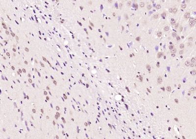
MAGI1 Polyclonal Antibody
Purified Rabbit Polyclonal Antibody (Pab)
- SPECIFICATION
- CITATIONS
- PROTOCOLS
- BACKGROUND

Application
| WB, IHC-P, IHC-F, IF, ICC, E |
|---|---|
| Primary Accession | Q96QZ7 |
| Reactivity | Rat, Pig, Dog, Bovine |
| Host | Rabbit |
| Clonality | Polyclonal |
| Calculated MW | 137 KDa |
| Physical State | Liquid |
| Immunogen | KLH conjugated synthetic peptide derived from human MAGI1 |
| Epitope Specificity | 161-260/1491 |
| Isotype | IgG |
| Purity | affinity purified by Protein A |
| Buffer | 0.01M TBS (pH7.4) with 1% BSA, 0.02% Proclin300 and 50% Glycerol. |
| SUBCELLULAR LOCATION | Cell junction; cell membrane; tight junction; peripheral membrane protein. Localizes to epithelial cells tight junctions. |
| SIMILARITY | Contains 1 guanylate kinase-like domain. Contains 6 PDZ (DHR) domains. Contains 2 WW domains. |
| SUBUNIT | Interacts through its WW 2 domain with SYNPO and through its PDZ 5 domain with ACTN4. Interacts with cytoplasmic domain of BAI1. Interacts via its WW domains with DRPLA. Interacts with ESAM, LRP2 and CXADR. May interact with CTNNB1. Interacts through its PDZ 1 domain with NET1 (By similarity). Interacts with ASIC3 and AMOT. Interacts with FCHSD2. Interacts with IGSF5/JAM4 and through its PDZ 2 and 3 domains with NPHS1 forming a tripartite complex (By similarity). Interacts with DDN. |
| Important Note | This product as supplied is intended for research use only, not for use in human, therapeutic or diagnostic applications. |
| Background Descriptions | The membrane-associated guanylate kinase (MAGUK) proteins are concentrated at the membrane-cytoskeletal interface where they facilitate the assembly of multiprotein complexes on the inner surface of the plasma membrane. Three protein-protein interaction modules characteristically define MAGUK related proteins: the PDZ domain, the SH3 domain and the guanylate kinase (GuK) domain. The closely related MAGUK proteins, MAGI-1, MAGI-2 and MAGI-3 (membrane associated guanylate kinase inverted-1 and 2), likewise contain the GuK domain and five PDZ domains; however, the SH3 domain is replaced with a WW domain. The transcripts of MAGI-1 are alternatively spliced to produce three distinct proteins having unique C-terminals. Two variants, MAGI-1a and MAGI-1b, are associated with the membrane and cytosolic fractions and are primarily expressed in the brain. The third isoform, MAGI-1c, encodes for a nuclear localization signal that localizes MAGI-1c to the nucleus, and it is primarily expressed in the liver and kidney. MAGI-2 and MAGI-3 are localized to the plasma membrane, and they contribute to protein scaffolding by associating with the protein phosphatase PTEN. |
| Gene ID | 9223 |
|---|---|
| Other Names | Membrane-associated guanylate kinase, WW and PDZ domain-containing protein 1, Atrophin-1-interacting protein 3, AIP-3, BAI1-associated protein 1, BAP-1, Membrane-associated guanylate kinase inverted 1, MAGI-1, Trinucleotide repeat-containing gene 19 protein, WW domain-containing protein 3, WWP3, MAGI1, AIP3, BAIAP1, BAP1, TNRC19 |
| Target/Specificity | Widely expressed with the exception of skeletal muscle. Isoform 1, isoform 2 and isoform 6 are highly expressed in colon, kidney, lung, liver, and pancreas. Isoform 5 is predominantly expressed in brain and heart. Isoform 3 and isoform 4 are highly expressed in pancreas and brain. |
| Dilution | WB=1:500-2000,IHC-P=1:100-500,IHC-F=1:100-500,ICC=1:100-500,IF=1:100-500,ELISA=1:5000-10000 |
| Storage | Store at -20 ℃ for one year. Avoid repeated freeze/thaw cycles. When reconstituted in sterile pH 7.4 0.01M PBS or diluent of antibody the antibody is stable for at least two weeks at 2-4 ℃. |
| Name | MAGI1 |
|---|---|
| Synonyms | AIP3, BAIAP1, BAP1, TNRC19 |
| Function | Plays a role in coupling actin fibers to cell junctions in endothelial cells, via its interaction with AMOTL2 and CDH5 (By similarity). May regulate acid-induced ASIC3 currents by modulating its expression at the cell surface (By similarity). |
| Cellular Location | Cell junction, tight junction. Cell membrane; Peripheral membrane protein. Note=Localizes to epithelial cells tight junctions |
| Tissue Location | Widely expressed with the exception of skeletal muscle. Isoform 1, isoform 2 and isoform 6 are highly expressed in colon, kidney, lung, liver, and pancreas. Isoform 5 is predominantly expressed in brain and heart. Isoform 3 and isoform 4 are highly expressed in pancreas and brain. |
Research Areas
Citations (0)

Thousands of laboratories across the world have published research that depended on the performance of antibodies from Abcepta to advance their research. Check out links to articles that cite our products in major peer-reviewed journals, organized by research category.
Submit your citation using an Abcepta antibody to
info@abcepta.com, and receive a free "I Love Antibodies" mug.
info@abcepta.com, and receive a free "I Love Antibodies" mug.

Application Protocols
Provided below are standard protocols that you may find useful for product applications.
Abcepta welcomes feedback from its customers.
If you have used an Abcepta product and would like to share how it has performed, please click on the "Submit Review" button and provide the requested information. Our staff will examine and post your review and contact you if needed.
If you have any additional inquiries please email technical services at tech@abcepta.com.
$ 385.00
Cat# AP54467
Ordering Information
United States
AlbaniaAustraliaAustriaBelgiumBosnia & HerzegovinaBrazilBulgariaCanadaCentral AmericaChinaCroatiaCyprusCzech RepublicDenmarkEstoniaFinlandFranceGermanyGreeceHong KongHungaryIcelandIndiaIndonesiaIrelandIsraelItalyJapanLatviaLithuaniaLuxembourgMacedoniaMalaysiaMaltaMexicoNetherlandsNew ZealandNorwayPakistanPolandPortugalRomaniaSerbiaSingaporeSlovakiaSloveniaSouth AfricaSouth KoreaSpainSwedenSwitzerlandTaiwanTurkeyUnited KingdomUnited StatesVietnamWorldwideOthers
USA Headquarters
(888) 735-7227 / (858) 622-0099 or (858) 875-1900
Shipping Information
Domestic orders (in stock items)
Shipped out the same day. Orders placed after 1 PM (PST) will ship out the next business day.
International orders
Contact your local distributors
