


 Foundational characteristics of cancer include proliferation, angiogenesis, migration, evasion of apoptosis, and cellular immortality. Find key markers for these cellular processes and antibodies to detect them.
 The SUMOplot™ Analysis Program predicts and scores sumoylation sites in your protein. SUMOylation is a post-translational modification involved in various cellular processes, such as nuclear-cytosolic transport, transcriptional regulation, apoptosis, protein stability, response to stress, and progression through the cell cycle.
 The Autophagy Receptor Motif Plotter predicts and scores autophagy receptor binding sites in your protein. Identifying proteins connected to this pathway is critical to understanding the role of autophagy in physiological as well as pathological processes such as development, differentiation, neurodegenerative diseases, stress, infection, and cancer.
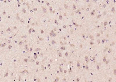
Advillin Polyclonal Antibody
Purified Rabbit Polyclonal Antibody (Pab)
- SPECIFICATION
- CITATIONS
- PROTOCOLS
- BACKGROUND

Application
| WB, IHC-P, IHC-F, IF, ICC, E |
|---|---|
| Primary Accession | O75366 |
| Reactivity | Rat, Pig, Dog, Bovine |
| Host | Rabbit |
| Clonality | Polyclonal |
| Calculated MW | 90 KDa |
| Physical State | Liquid |
| Immunogen | KLH conjugated synthetic peptide derived from human Advillin |
| Epitope Specificity | 151-250/819 |
| Isotype | IgG |
| Purity | affinity purified by Protein A |
| Buffer | 0.01M TBS (pH7.4) with 1% BSA, 0.02% Proclin300 and 50% Glycerol. |
| SUBCELLULAR LOCATION | Cytoplasm > cytoskeleton. Cell projection. Cell projection > axon. |
| SIMILARITY | Belongs to the villin/gelsolin family. Contains 6 gelsolin-like repeats. Contains 1 HP (headpiece) domain. |
| SUBUNIT | Associates (via C-terminus) with actin. Interacts with SCARF1 (By similarity). Interacts with F-actin. |
| Important Note | This product as supplied is intended for research use only, not for use in human, therapeutic or diagnostic applications. |
| Background Descriptions | Advillin is an 819 amino acid protein that localizes to both the cytoplasm and the cytoskeleton and contains one HP domain and six gelsolin-like repeats. Expressed at high levels in colon and small intestine and at lower levels in uterus, thymus, testis and prostate, advillin functions as a calcium-regulated Actin-binding protein that may be involved in the development of neuronal cells, specifically those that form ganglia. The gene encoding advillin maps to human chromosome 12, which encodes over 1,100 genes and comprises approximately 4.5% of the human genome. Chromosome 12 is associated with a variety of diseases and afflictions, including hypochondrogenesis, achondrogenesis, Kniest dysplasia, Noonan syndrome and Trisomy 12p, which causes facial developmental defects and seizure disorders. |
| Gene ID | 10677 |
|---|---|
| Other Names | Advillin, p92, AVIL (HGNC:14188) |
| Target/Specificity | Most highly expressed in the small intestine and colonic lining. Weaker expression also detected in the thymus, prostate, testes and uterus. |
| Dilution | WB=1:500-2000,IHC-P=1:100-500,IHC-F=1:100-500,ICC=1:100-500,IF=1:100-500,ELISA=1:5000-10000 |
| Storage | Store at -20 ℃ for one year. Avoid repeated freeze/thaw cycles. When reconstituted in sterile pH 7.4 0.01M PBS or diluent of antibody the antibody is stable for at least two weeks at 2-4 ℃. |
| Name | AVIL (HGNC:14188) |
|---|---|
| Function | Ca(2+)-regulated actin-binding protein which plays an important role in actin bundling (PubMed:29058690). May have a unique function in the morphogenesis of neuronal cells which form ganglia. Required for SREC1-mediated regulation of neurite-like outgrowth. Plays a role in regenerative sensory axon outgrowth and remodeling processes after peripheral injury in neonates. Involved in the formation of long fine actin-containing filopodia-like structures in fibroblast. Plays a role in ciliogenesis. In podocytes, controls lamellipodia formation through the regulation of EGF-induced diacylglycerol generation by PLCE1 and ARP2/3 complex assembly (PubMed:29058690). |
| Cellular Location | Cytoplasm, cytoskeleton. Cell projection, lamellipodium. Cell junction, focal adhesion. Cell projection, neuron projection {ECO:0000250|UniProtKB:Q9WU06}. Cell projection, axon {ECO:0000250|UniProtKB:Q9WU06}. Note=In podocytes, present in the F- actin-enriched cell periphery that generates lamellipodia and focal adhesions. |
| Tissue Location | Most highly expressed in the small intestine and colonic lining. Weaker expression also detected in the thymus, prostate, testes and uterus (PubMed:12034507). Expressed in podocytes (at protein level) (PubMed:29058690). |
Research Areas
Citations (0)

Thousands of laboratories across the world have published research that depended on the performance of antibodies from Abcepta to advance their research. Check out links to articles that cite our products in major peer-reviewed journals, organized by research category.
Submit your citation using an Abcepta antibody to
info@abcepta.com, and receive a free "I Love Antibodies" mug.
info@abcepta.com, and receive a free "I Love Antibodies" mug.

Application Protocols
Provided below are standard protocols that you may find useful for product applications.
Abcepta welcomes feedback from its customers.
If you have used an Abcepta product and would like to share how it has performed, please click on the "Submit Review" button and provide the requested information. Our staff will examine and post your review and contact you if needed.
If you have any additional inquiries please email technical services at tech@abcepta.com.
$ 385.00
Cat# AP54508
Ordering Information
United States
AlbaniaAustraliaAustriaBelgiumBosnia & HerzegovinaBrazilBulgariaCanadaCentral AmericaChinaCroatiaCyprusCzech RepublicDenmarkEstoniaFinlandFranceGermanyGreeceHong KongHungaryIcelandIndiaIndonesiaIrelandIsraelItalyJapanLatviaLithuaniaLuxembourgMacedoniaMalaysiaMaltaMexicoNetherlandsNew ZealandNorwayPakistanPolandPortugalRomaniaSerbiaSingaporeSlovakiaSloveniaSouth AfricaSouth KoreaSpainSwedenSwitzerlandTaiwanTurkeyUnited KingdomUnited StatesVietnamWorldwideOthers
USA Headquarters
(888) 735-7227 / (858) 622-0099 or (858) 875-1900
Shipping Information
Domestic orders (in stock items)
Shipped out the same day. Orders placed after 1 PM (PST) will ship out the next business day.
International orders
Contact your local distributors
