


 Foundational characteristics of cancer include proliferation, angiogenesis, migration, evasion of apoptosis, and cellular immortality. Find key markers for these cellular processes and antibodies to detect them.
 The SUMOplot™ Analysis Program predicts and scores sumoylation sites in your protein. SUMOylation is a post-translational modification involved in various cellular processes, such as nuclear-cytosolic transport, transcriptional regulation, apoptosis, protein stability, response to stress, and progression through the cell cycle.
 The Autophagy Receptor Motif Plotter predicts and scores autophagy receptor binding sites in your protein. Identifying proteins connected to this pathway is critical to understanding the role of autophagy in physiological as well as pathological processes such as development, differentiation, neurodegenerative diseases, stress, infection, and cancer.
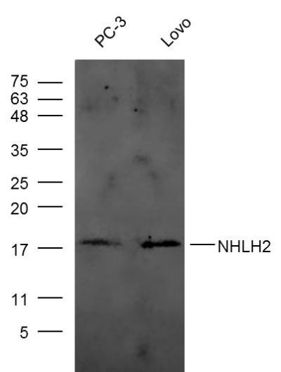
NHLH2 Polyclonal Antibody
Purified Rabbit Polyclonal Antibody (Pab)
- SPECIFICATION
- CITATIONS
- PROTOCOLS
- BACKGROUND

Application
| WB, IHC-P, IHC-F, IF, ICC, E |
|---|---|
| Primary Accession | Q02577 |
| Reactivity | Rat, Pig, Dog, Bovine |
| Host | Rabbit |
| Clonality | Polyclonal |
| Calculated MW | 15 KDa |
| Physical State | Liquid |
| Immunogen | KLH conjugated synthetic peptide derived from human NHLH2 |
| Epitope Specificity | 1-50/135 |
| Isotype | IgG |
| Purity | affinity purified by Protein A |
| Buffer | 0.01M TBS (pH7.4) with 1% BSA, 0.02% Proclin300 and 50% Glycerol. |
| SUBCELLULAR LOCATION | Nucleus |
| SIMILARITY | Contains 1 bHLH (basic helix-loop-helix) domain. |
| SUBUNIT | Efficient DNA binding requires dimerization with another bHLH protein. |
| Important Note | This product as supplied is intended for research use only, not for use in human, therapeutic or diagnostic applications. |
| Background Descriptions | The helix-loop-helix (HLH) structures are known motifs commonly found in membrane-active and DNA-binding proteins. The helix-loop-helix proteins HEN1 and HEN2 are DNA-binding proteins that may be involved in cell-type determination in the early nervous system. Studies of expression in normal tissues have demonstrated expression of NHLH1/NSCL-1 and NHLH2/NSCL-2, the genes encoding HEN1 and HEN2, in the developing central and peripheral nervous system, specifically in developing neurons. |
| Gene ID | 4808 |
|---|---|
| Other Names | Helix-loop-helix protein 2, HEN-2, Class A basic helix-loop-helix protein 34, bHLHa34, Nescient helix loop helix 2, NSCL-2, NHLH2, BHLHA34, HEN2, KIAA0490 |
| Dilution | WB=1:500-2000,IHC-P=1:100-500,IHC-F=1:100-500,ICC=1:100-500,IF=1:100-500,ELISA=1:5000-10000 |
| Storage | Store at -20 ℃ for one year. Avoid repeated freeze/thaw cycles. When reconstituted in sterile pH 7.4 0.01M PBS or diluent of antibody the antibody is stable for at least two weeks at 2-4 ℃. |
| Name | NHLH2 |
|---|---|
| Synonyms | BHLHA34, HEN2, KIAA0490 |
| Function | Transcription factor which binds the E box motif 5'- CA[TC][AG]TG-3'. Involved in regulating energy expenditure, body mass, voluntary physical activity, mating behavior and reproductive longevity, acting through the hypothalamic-pituitary-gonadal axis. Acts as a transcriptional activator of target genes, including NDN, PCSK1, MC4R (By similarity). Is also a transcriptional activator of KISS1 (PubMed:35066646). May act centrally to regulate function of both white and brown adipose tissue. Together with NHLH1, required to maintain migration and survival of cells in the anterior extramural migration stream (aes), which forms the precerebellar nuclei. Also, in concert with NHLH1, may determine fate of gonadotropin releasing hormone-1 (GnRH-1) neurons. |
| Cellular Location | Nucleus {ECO:0000255|PROSITE-ProRule:PRU00981}. |
Research Areas
Citations (0)

Thousands of laboratories across the world have published research that depended on the performance of antibodies from Abcepta to advance their research. Check out links to articles that cite our products in major peer-reviewed journals, organized by research category.
Submit your citation using an Abcepta antibody to
info@abcepta.com, and receive a free "I Love Antibodies" mug.
info@abcepta.com, and receive a free "I Love Antibodies" mug.

Application Protocols
Provided below are standard protocols that you may find useful for product applications.
Abcepta welcomes feedback from its customers.
If you have used an Abcepta product and would like to share how it has performed, please click on the "Submit Review" button and provide the requested information. Our staff will examine and post your review and contact you if needed.
If you have any additional inquiries please email technical services at tech@abcepta.com.
$ 385.00
Cat# AP54532
Ordering Information
United States
AlbaniaAustraliaAustriaBelgiumBosnia & HerzegovinaBrazilBulgariaCanadaCentral AmericaChinaCroatiaCyprusCzech RepublicDenmarkEstoniaFinlandFranceGermanyGreeceHong KongHungaryIcelandIndiaIndonesiaIrelandIsraelItalyJapanLatviaLithuaniaLuxembourgMacedoniaMalaysiaMaltaMexicoNetherlandsNew ZealandNorwayPakistanPolandPortugalRomaniaSerbiaSingaporeSlovakiaSloveniaSouth AfricaSouth KoreaSpainSwedenSwitzerlandTaiwanTurkeyUnited KingdomUnited StatesVietnamWorldwideOthers
USA Headquarters
(888) 735-7227 / (858) 622-0099 or (858) 875-1900
Other Products
Shipping Information
Domestic orders (in stock items)
Shipped out the same day. Orders placed after 1 PM (PST) will ship out the next business day.
International orders
Contact your local distributors
