


 Foundational characteristics of cancer include proliferation, angiogenesis, migration, evasion of apoptosis, and cellular immortality. Find key markers for these cellular processes and antibodies to detect them.
 The SUMOplot™ Analysis Program predicts and scores sumoylation sites in your protein. SUMOylation is a post-translational modification involved in various cellular processes, such as nuclear-cytosolic transport, transcriptional regulation, apoptosis, protein stability, response to stress, and progression through the cell cycle.
 The Autophagy Receptor Motif Plotter predicts and scores autophagy receptor binding sites in your protein. Identifying proteins connected to this pathway is critical to understanding the role of autophagy in physiological as well as pathological processes such as development, differentiation, neurodegenerative diseases, stress, infection, and cancer.
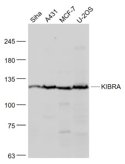
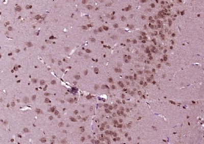
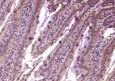
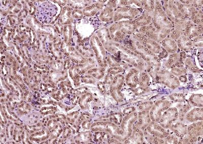
KIBRA Polyclonal Antibody
Purified Rabbit Polyclonal Antibody (Pab)
- SPECIFICATION
- CITATIONS
- PROTOCOLS
- BACKGROUND

Application
| WB, IHC-P, IHC-F, IF, ICC, E |
|---|---|
| Primary Accession | Q8IX03 |
| Reactivity | Rat, Dog, Bovine |
| Host | Rabbit |
| Clonality | Polyclonal |
| Calculated MW | 125 KDa |
| Physical State | Liquid |
| Immunogen | KLH conjugated synthetic peptide derived from human KIBRA |
| Epitope Specificity | 351-450/1113 |
| Isotype | IgG |
| Purity | affinity purified by Protein A |
| Buffer | 0.01M TBS (pH7.4) with 1% BSA, 0.02% Proclin300 and 50% Glycerol. |
| SUBCELLULAR LOCATION | Cytoplasm. Cytoplasm, perinuclear region. Nucleus. Cell projection, ruffle membrane. Note=Co-localizes with PRKCZ in the perinuclear region. |
| SIMILARITY | Belongs to the WWC family. KIBRA subfamily. Contains 1 C2 domain. Contains 2 WW domains. |
| SUBUNIT | Homodimer. Interacts with DDN. Interacts with DYNLL1 and histone H3. The interaction with DYNLL1 is mandatory for the recruitment and transactivation functions of ESR1 or DYNLL1 to the target chromatin and the interaction with histone H3 ensures proper regulatory interaction of WWC1-DYNLL1-ESR1 complexes with target chromatin. Interacts (via WW domains) with DDR1 (via PPxY motif) in a collagen-regulated manner. Interacts with PRKCZ (via the protein kinase domain). Forms a tripartite complex with DDR1 and PRKCZ, but predominantly in the absence of collagen. Interacts (via the ADDV motif) with INADL (via PDZ domain 8). Interacts (via WW domains) with SYNPO (via PPxY motifs). Interacts with NF2 and SNX4. |
| Important Note | This product as supplied is intended for research use only, not for use in human, therapeutic or diagnostic applications. |
| Background Descriptions | KIBRA is a 1,113 amino acid protein that localizes to the cytoplasm and contains one C2 domain and two WW domains. Expressed in colon, brain, kidney and heart tissue, KIBRA is thought to interact with dendrin and, via this interaction, may play a role in collagen-induced signaling. Additionally, KIBRA, which exists as multiple alternatively spliced isoforms, is involved in memory performance and in the pathogenesis of Alzheimer's disease. The gene encoding KIBRA maps to human chromosome 5, which contains 181 million base pairs and comprises nearly 6% of the human genome. Deletion of the p arm of chromosome 5 leads to Cri du chat syndrome, while deletion of the q arm or of chromosome 5 altogether is common in therapy-related acute myelogenous leukemias and myelodysplastic syndrome. |
| Gene ID | 23286 |
|---|---|
| Other Names | Protein KIBRA, HBeAg-binding protein 3, Kidney and brain protein, KIBRA, WW domain-containing protein 1, WWC1, KIAA0869 |
| Target/Specificity | Expressed in mammary epithelial cells and breast cancer cell lines. Found in the luminal epithelium surrounding the ducts in the normal breast. In the brain, expressed in somatodendritic compartment of neurons in the cortex and hippocampus and in the cerebellum it is found in the Purkinje cells and some granule cells (at protein level). Detected in brain, heart, colon and kidney. In the kidney, expressed in glomerular podocytes, in some tubules and in the collecting duct. |
| Dilution | WB=1:500-2000,IHC-P=1:100-500,IHC-F=1:100-500,ICC=1:100-500,IF=1:100-500,ELISA=1:5000-10000 |
| Storage | Store at -20 ℃ for one year. Avoid repeated freeze/thaw cycles. When reconstituted in sterile pH 7.4 0.01M PBS or diluent of antibody the antibody is stable for at least two weeks at 2-4 ℃. |
| Name | WWC1 (HGNC:29435) |
|---|---|
| Synonyms | KIAA0869, KIBRA |
| Function | Regulator of the Hippo signaling pathway, also known as the Salvador-Warts-Hippo (SWH) pathway (PubMed:24682284). Enhances phosphorylation of LATS1 and YAP1 and negatively regulates cell proliferation and organ growth due to a suppression of the transcriptional activity of YAP1, the major effector of the Hippo pathway (PubMed:24682284). Along with NF2 can synergistically induce the phosphorylation of LATS1 and LATS2 and function in the regulation of Hippo signaling pathway (PubMed:20159598). Acts as a transcriptional coactivator of ESR1 which plays an essential role in DYNLL1-mediated ESR1 transactivation (PubMed:16684779). Regulates collagen-stimulated activation of the ERK/MAPK cascade (PubMed:18190796). Modulates directional migration of podocytes (PubMed:18596123). Plays a role in cognition and memory performance (PubMed:18672031). Plays an important role in regulating AMPA-selective glutamate receptors (AMPARs) trafficking underlying synaptic plasticity and learning (By similarity). |
| Cellular Location | Cytoplasm, perinuclear region. Nucleus. Cell projection, ruffle membrane. Cytoplasm, cytosol. Note=Colocalizes with PRKCZ in the perinuclear region |
| Tissue Location | Expressed in mammary epithelial cells and breast cancer cell lines. Found in the luminal epithelium surrounding the ducts in the normal breast. In the brain, expressed in somatodendritic compartment of neurons in the cortex and hippocampus and in the cerebellum it is found in the Purkinje cells and some granule cells (at protein level). Detected in brain, heart, colon and kidney. In the kidney, expressed in glomerular podocytes, in some tubules and in the collecting duct. |
Research Areas
Citations (0)

Thousands of laboratories across the world have published research that depended on the performance of antibodies from Abcepta to advance their research. Check out links to articles that cite our products in major peer-reviewed journals, organized by research category.
Submit your citation using an Abcepta antibody to
info@abcepta.com, and receive a free "I Love Antibodies" mug.
info@abcepta.com, and receive a free "I Love Antibodies" mug.

Application Protocols
Provided below are standard protocols that you may find useful for product applications.
Abcepta welcomes feedback from its customers.
If you have used an Abcepta product and would like to share how it has performed, please click on the "Submit Review" button and provide the requested information. Our staff will examine and post your review and contact you if needed.
If you have any additional inquiries please email technical services at tech@abcepta.com.
$ 385.00
Cat# AP54550
Ordering Information
United States
AlbaniaAustraliaAustriaBelgiumBosnia & HerzegovinaBrazilBulgariaCanadaCentral AmericaChinaCroatiaCyprusCzech RepublicDenmarkEstoniaFinlandFranceGermanyGreeceHong KongHungaryIcelandIndiaIndonesiaIrelandIsraelItalyJapanLatviaLithuaniaLuxembourgMacedoniaMalaysiaMaltaMexicoNetherlandsNew ZealandNorwayPakistanPolandPortugalRomaniaSerbiaSingaporeSlovakiaSloveniaSouth AfricaSouth KoreaSpainSwedenSwitzerlandTaiwanTurkeyUnited KingdomUnited StatesVietnamWorldwideOthers
USA Headquarters
(888) 735-7227 / (858) 622-0099 or (858) 875-1900
Other Products
Shipping Information
Domestic orders (in stock items)
Shipped out the same day. Orders placed after 1 PM (PST) will ship out the next business day.
International orders
Contact your local distributors
