


 Foundational characteristics of cancer include proliferation, angiogenesis, migration, evasion of apoptosis, and cellular immortality. Find key markers for these cellular processes and antibodies to detect them.
 The SUMOplot™ Analysis Program predicts and scores sumoylation sites in your protein. SUMOylation is a post-translational modification involved in various cellular processes, such as nuclear-cytosolic transport, transcriptional regulation, apoptosis, protein stability, response to stress, and progression through the cell cycle.
 The Autophagy Receptor Motif Plotter predicts and scores autophagy receptor binding sites in your protein. Identifying proteins connected to this pathway is critical to understanding the role of autophagy in physiological as well as pathological processes such as development, differentiation, neurodegenerative diseases, stress, infection, and cancer.
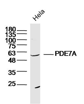
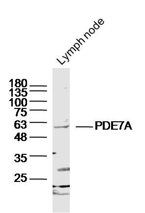
PDE7A Polyclonal Antibody
Purified Rabbit Polyclonal Antibody (Pab)
- SPECIFICATION
- CITATIONS
- PROTOCOLS
- BACKGROUND

Application
| WB, IHC-P, IHC-F, IF, ICC, E |
|---|---|
| Primary Accession | Q13946 |
| Reactivity | Rat, Pig, Dog, Bovine |
| Host | Rabbit |
| Clonality | Polyclonal |
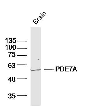
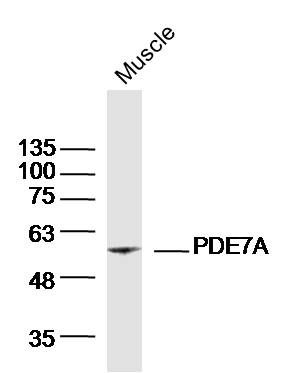
| Calculated MW | 55 KDa |
| Physical State | Liquid |
| Immunogen | KLH conjugated synthetic peptide derived from human PDE7A |
| Epitope Specificity | 341-440/482 |
| Isotype | IgG |
| Purity | affinity purified by Protein A |
| Buffer | 0.01M TBS (pH7.4) with 1% BSA, 0.02% Proclin300 and 50% Glycerol. |
| SUBCELLULAR LOCATION | Cytoplasm |
| SIMILARITY | Belongs to the cyclic nucleotide phosphodiesterase family. PDE7 subfamily. |
| SUBUNIT | Interacts with CBFA2T3. |
| Important Note | This product as supplied is intended for research use only, not for use in human, therapeutic or diagnostic applications. |
| Background Descriptions | Phosphodiesterases (PDE, also designated cyclic nucleotide phosphodiesterase) are important for the downregulation of the intracellular level of the second messenger cyclic adenosine monophosphate (cAMP) by hydrolyzing cAMP to 5'AMP. Phosphodiesterase type 3 isoforms, PDE3A and 3B, are expressed primarily in cardiovascular tissue and adipose tissue, respectively. PDE3A, is found in myocardium and platelets and PDE3B is found in lymphocytes. The PDE7A1 (HCP1) isozyme and the PDE7A2 proteins, alternate splice products of PDE7A, are highly expressed in skeletal muscle. PDE7B is most highly expressed in pancreas. The PDE family contains proteins that serve tissue-specific roles in regulation of lipolysis, glycogenolysis, myocardial contractility, and smooth muscle relaxation. |
| Gene ID | 5150 |
|---|---|
| Other Names | High affinity cAMP-specific 3', 5'-cyclic phosphodiesterase 7A, 3.1.4.53, HCP1, TM22, PDE7A |
| Target/Specificity | PDE7A1 is found at high levels in skeletal muscle and at low levels in a variety of tissues including brain and heart. It is expressed as well in two T-cell lines. PDE7A2 is found abundantly in skeletal muscle and at low levels in heart. |
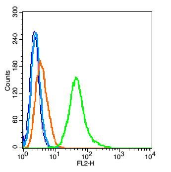
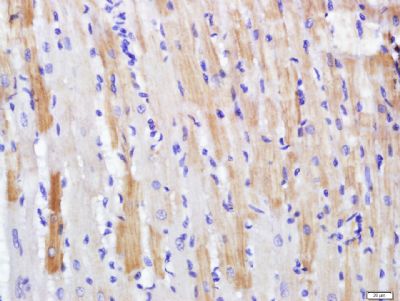
| Dilution | WB=1:500-2000,IHC-P=1:100-500,IHC-F=1:100-500,ICC=1:100-500,IF=1:100-500,Flow-Cyt=1 µg/Test,ELISA=1:5000-10000 |
| Storage | Store at -20 ℃ for one year. Avoid repeated freeze/thaw cycles. When reconstituted in sterile pH 7.4 0.01M PBS or diluent of antibody the antibody is stable for at least two weeks at 2-4 ℃. |
| Name | PDE7A {ECO:0000303|PubMed:9195912, ECO:0000312|HGNC:HGNC:8791} |
|---|---|
| Function | Hydrolyzes the second messenger cAMP, which is a key regulator of many important physiological processes (PubMed:19350606, PubMed:8389765, PubMed:9195912). May have a role in muscle signal transduction (PubMed:9195912). |
| Cellular Location | [Isoform PDE7A1]: Cytoplasm, cytosol. Note=PDE7A1 (57 kDa) is located mostly to soluble cellular fractions. |
| Tissue Location | [Isoform PDE7A1]: Found at high levels in skeletal muscle and at low levels in a variety of tissues including brain and heart (PubMed:9195912). It is expressed as well in two T-cell lines (PubMed:9195912). |
Research Areas
Citations (0)

Thousands of laboratories across the world have published research that depended on the performance of antibodies from Abcepta to advance their research. Check out links to articles that cite our products in major peer-reviewed journals, organized by research category.
Submit your citation using an Abcepta antibody to
info@abcepta.com, and receive a free "I Love Antibodies" mug.
info@abcepta.com, and receive a free "I Love Antibodies" mug.

Application Protocols
Provided below are standard protocols that you may find useful for product applications.
Abcepta welcomes feedback from its customers.
If you have used an Abcepta product and would like to share how it has performed, please click on the "Submit Review" button and provide the requested information. Our staff will examine and post your review and contact you if needed.
If you have any additional inquiries please email technical services at tech@abcepta.com.
$ 385.00
Cat# AP54552
Ordering Information
United States
AlbaniaAustraliaAustriaBelgiumBosnia & HerzegovinaBrazilBulgariaCanadaCentral AmericaChinaCroatiaCyprusCzech RepublicDenmarkEstoniaFinlandFranceGermanyGreeceHong KongHungaryIcelandIndiaIndonesiaIrelandIsraelItalyJapanLatviaLithuaniaLuxembourgMacedoniaMalaysiaMaltaMexicoNetherlandsNew ZealandNorwayPakistanPolandPortugalRomaniaSerbiaSingaporeSlovakiaSloveniaSouth AfricaSouth KoreaSpainSwedenSwitzerlandTaiwanTurkeyUnited KingdomUnited StatesVietnamWorldwideOthers
USA Headquarters
(888) 735-7227 / (858) 622-0099 or (858) 875-1900
Shipping Information
Domestic orders (in stock items)
Shipped out the same day. Orders placed after 1 PM (PST) will ship out the next business day.
International orders
Contact your local distributors
