


 Foundational characteristics of cancer include proliferation, angiogenesis, migration, evasion of apoptosis, and cellular immortality. Find key markers for these cellular processes and antibodies to detect them.
 The SUMOplot™ Analysis Program predicts and scores sumoylation sites in your protein. SUMOylation is a post-translational modification involved in various cellular processes, such as nuclear-cytosolic transport, transcriptional regulation, apoptosis, protein stability, response to stress, and progression through the cell cycle.
 The Autophagy Receptor Motif Plotter predicts and scores autophagy receptor binding sites in your protein. Identifying proteins connected to this pathway is critical to understanding the role of autophagy in physiological as well as pathological processes such as development, differentiation, neurodegenerative diseases, stress, infection, and cancer.
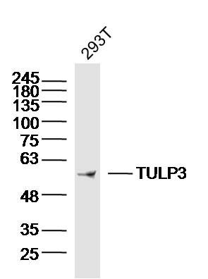
TULP3 Polyclonal Antibody
Purified Rabbit Polyclonal Antibody (Pab)
- SPECIFICATION
- CITATIONS
- PROTOCOLS
- BACKGROUND

Application
| WB, IHC-P, IHC-F, IF, ICC, E |
|---|---|
| Primary Accession | O75386 |
| Reactivity | Rat, Dog |
| Host | Rabbit |
| Clonality | Polyclonal |
| Calculated MW | 50 KDa |
| Physical State | Liquid |
| Immunogen | KLH conjugated synthetic peptide derived from human TULP3 |
| Epitope Specificity | 251-350/442 |
| Isotype | IgG |
| Purity | affinity purified by Protein A |
| Buffer | 0.01M TBS (pH7.4) with 1% BSA, 0.02% Proclin300 and 50% Glycerol. |
| SUBCELLULAR LOCATION | Nucleus. Cell membrane. Cell projection, cilium (By similarity). Cytoplasm (By similarity). Secreted (By similarity). Note=Does not have a cleavable signal peptide and is secreted by a non-conventional pathway (By similarity). Translocates from the plasma membrane to the nucleus upon activation of guanine nucleotide-binding protein G(q) subunit alpha. |
| SIMILARITY | Belongs to the TUB family. |
| Important Note | This product as supplied is intended for research use only, not for use in human, therapeutic or diagnostic applications. |
| Background Descriptions | Mutations in the mouse Tub gene gradually lead to obesity, strongly resembling the late-onset obesity observed in the human population. In addition to excessive deposition of adipose tissue, mice with the Tub phenotype also suffer retinal degeneration and neurosensory hearing loss. A human homolog of the Tub gene has been identified, as have three related proteins, called Tubby-like protein 1 (TULP1), TULP2 and TULP3. When compared to TULP1 and TULP2, TULP3 has a wider tissue expression and is phylogenetically more similar to Tub than either TULP1 or TULP2. TULP1, expressed specifically in the retina, maps to the chromosomal region known to be involved in retinitis pigmentosa, while TULP2 maps within the minimal interval for the rod-cone dystrophy. TULP3 maps to human chromosome 12p13, and shares 69% homology to mouse TULP3. Human RNA from testis, ovary, thyroid and spinal cord contain highly detectable levels of TULP3 transcripts. In the retina, TULP3 is expressed specifically in the inner nuclear layer and ganglion cell layer. TULP1, TULP2 and TULP3 may comprise a unique family of bipartite transcription factors. |
| Gene ID | 7289 |
|---|---|
| Other Names | Tubby-related protein 3, Tubby-like protein 3, TULP3 (HGNC:12425), TUBL3 |
| Target/Specificity | Expressed at high levels in testis, ovaries, thyroid, and spinal chord. |
| Dilution | WB=1:500-2000,IHC-P=1:100-500,IHC-F=1:100-500,ICC=1:100-500,IF=1:100-500,ELISA=1:5000-10000 |
| Storage | Store at -20 ℃ for one year. Avoid repeated freeze/thaw cycles. When reconstituted in sterile pH 7.4 0.01M PBS or diluent of antibody the antibody is stable for at least two weeks at 2-4 ℃. |
| Name | TULP3 (HGNC:12425) |
|---|---|
| Synonyms | TUBL3 |
| Function | Negative regulator of the Shh signaling transduction pathway: recruited to primary cilia via association with the IFT complex A (IFT- A) and is required for recruitment of G protein-coupled receptor GPR161 to cilia, a promoter of PKA-dependent basal repression machinery in Shh signaling. Binds to phosphorylated inositide (phosphoinositide) lipids. Both IFT-A- and phosphoinositide-binding properties are required to regulate ciliary G protein-coupled receptor trafficking. During adipogenesis, regulates ciliary trafficking of FFAR4 in preadipocytes. |
| Cellular Location | Nucleus. Cell membrane. Cell projection, cilium. Cytoplasm. Secreted. Note=Does not have a cleavable signal peptide and is secreted by a non-conventional pathway (By similarity). Translocates from the plasma membrane to the nucleus upon activation of guanine nucleotide-binding protein G(q) subunit alpha |
| Tissue Location | Expressed at high levels in testis, ovaries, thyroid, and spinal cord. |
Research Areas
Citations (0)

Thousands of laboratories across the world have published research that depended on the performance of antibodies from Abcepta to advance their research. Check out links to articles that cite our products in major peer-reviewed journals, organized by research category.
Submit your citation using an Abcepta antibody to
info@abcepta.com, and receive a free "I Love Antibodies" mug.
info@abcepta.com, and receive a free "I Love Antibodies" mug.

Application Protocols
Provided below are standard protocols that you may find useful for product applications.
Abcepta welcomes feedback from its customers.
If you have used an Abcepta product and would like to share how it has performed, please click on the "Submit Review" button and provide the requested information. Our staff will examine and post your review and contact you if needed.
If you have any additional inquiries please email technical services at tech@abcepta.com.
$ 385.00
Cat# AP54565
Ordering Information
United States
AlbaniaAustraliaAustriaBelgiumBosnia & HerzegovinaBrazilBulgariaCanadaCentral AmericaChinaCroatiaCyprusCzech RepublicDenmarkEstoniaFinlandFranceGermanyGreeceHong KongHungaryIcelandIndiaIndonesiaIrelandIsraelItalyJapanLatviaLithuaniaLuxembourgMacedoniaMalaysiaMaltaMexicoNetherlandsNew ZealandNorwayPakistanPolandPortugalRomaniaSerbiaSingaporeSlovakiaSloveniaSouth AfricaSouth KoreaSpainSwedenSwitzerlandTaiwanTurkeyUnited KingdomUnited StatesVietnamWorldwideOthers
USA Headquarters
(888) 735-7227 / (858) 622-0099 or (858) 875-1900
Shipping Information
Domestic orders (in stock items)
Shipped out the same day. Orders placed after 1 PM (PST) will ship out the next business day.
International orders
Contact your local distributors
