


 Foundational characteristics of cancer include proliferation, angiogenesis, migration, evasion of apoptosis, and cellular immortality. Find key markers for these cellular processes and antibodies to detect them.
 The SUMOplot™ Analysis Program predicts and scores sumoylation sites in your protein. SUMOylation is a post-translational modification involved in various cellular processes, such as nuclear-cytosolic transport, transcriptional regulation, apoptosis, protein stability, response to stress, and progression through the cell cycle.
 The Autophagy Receptor Motif Plotter predicts and scores autophagy receptor binding sites in your protein. Identifying proteins connected to this pathway is critical to understanding the role of autophagy in physiological as well as pathological processes such as development, differentiation, neurodegenerative diseases, stress, infection, and cancer.
LRRC25 Polyclonal Antibody
Purified Rabbit Polyclonal Antibody (Pab)
- SPECIFICATION
- CITATIONS
- PROTOCOLS
- BACKGROUND

Application
| WB, IHC-P, IHC-F, IF, ICC, E |
|---|---|
| Primary Accession | Q8N386 |
| Host | Rabbit |
| Clonality | Polyclonal |
| Calculated MW | 31 KDa |
| Physical State | Liquid |
| Immunogen | KLH conjugated synthetic peptide derived from human LRRC25 |
| Epitope Specificity | 81-180/305 |
| Isotype | IgG |
| Purity | affinity purified by Protein A |
| Buffer | 0.01M TBS (pH7.4) with 1% BSA, 0.02% Proclin300 and 50% Glycerol. |
| SUBCELLULAR LOCATION | Membrane; Single-pass type I membrane protein. |
| Important Note | This product as supplied is intended for research use only, not for use in human, therapeutic or diagnostic applications. |
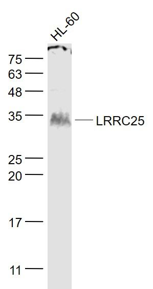
| Background Descriptions | LRRC25 is a 305 amino acid protein that contains two LRR repeats. LRRC25 is a single-pass type I membrane protein that is expressed in monocyte-derived dendritic cells (MDDC), granulocytes, monocytes, plasmacytoid dendritic cells (PDC), B-lymphocytes, peripheral blood leukocytes, spleen and bone marrow, with lower levels in lymph nodes, fetal liver, and appendix. LRRC25 may play a role in the activation of cells of innate and acquired immunity. |
| Gene ID | 126364 |
|---|---|
| Other Names | Leucine-rich repeat-containing protein 25, Monocyte and plasmacytoid-activated protein, LRRC25, MAPA |
| Target/Specificity | Expressed in plasmacytoid dendritic cells (PDC), monocyte-derived dendritic cells (MDDC), granulocytes, monocytes, B-lymphocytes, peripheral blood leukocytes, spleen, bone marrow, and, to a lesser extent, lymph nodes, fetal liver, and appendix but not in thymus. |
| Dilution | WB=1:500-2000,IHC-P=1:100-500,IHC-F=1:100-500,ICC=1:100-500,IF=1:100-500,ELISA=1:5000-10000 |
| Storage | Store at -20 ℃ for one year. Avoid repeated freeze/thaw cycles. When reconstituted in sterile pH 7.4 0.01M PBS or diluent of antibody the antibody is stable for at least two weeks at 2-4 ℃. |
| Name | LRRC25 |
|---|---|
| Synonyms | MAPA |
| Function | Plays a role in the inhibition of RLR-mediated type I interferon signaling pathway by targeting RIGI for autophagic degradation. Interacts specifically with ISG15-associated RIGI to promote interaction between RIGI and the autophagic cargo receptor p62/SQSTM1 to mediate RIGI degradation via selective autophagy (PubMed:29288164). Also plays a role in the inhibition of NF-kappa-B signaling pathway and inflammatory response by promoting the degradation of p65/RELA. |
| Cellular Location | Membrane; Single-pass type I membrane protein. Cytoplasm |
| Tissue Location | Expressed in plasmacytoid dendritic cells (PDC), monocyte-derived dendritic cells (MDDC), granulocytes, monocytes, B- lymphocytes, peripheral blood leukocytes, spleen, bone marrow, and, to a lesser extent, lymph nodes, fetal liver, and appendix but not in thymus. |
Research Areas
Citations (0)

Thousands of laboratories across the world have published research that depended on the performance of antibodies from Abcepta to advance their research. Check out links to articles that cite our products in major peer-reviewed journals, organized by research category.
Submit your citation using an Abcepta antibody to
info@abcepta.com, and receive a free "I Love Antibodies" mug.
info@abcepta.com, and receive a free "I Love Antibodies" mug.

Application Protocols
Provided below are standard protocols that you may find useful for product applications.
Abcepta welcomes feedback from its customers.
If you have used an Abcepta product and would like to share how it has performed, please click on the "Submit Review" button and provide the requested information. Our staff will examine and post your review and contact you if needed.
If you have any additional inquiries please email technical services at tech@abcepta.com.
$ 385.00
Cat# AP54829
Ordering Information
United States
AlbaniaAustraliaAustriaBelgiumBosnia & HerzegovinaBrazilBulgariaCanadaCentral AmericaChinaCroatiaCyprusCzech RepublicDenmarkEstoniaFinlandFranceGermanyGreeceHong KongHungaryIcelandIndiaIndonesiaIrelandIsraelItalyJapanLatviaLithuaniaLuxembourgMacedoniaMalaysiaMaltaMexicoNetherlandsNew ZealandNorwayPakistanPolandPortugalRomaniaSerbiaSingaporeSlovakiaSloveniaSouth AfricaSouth KoreaSpainSwedenSwitzerlandTaiwanTurkeyUnited KingdomUnited StatesVietnamWorldwideOthers
USA Headquarters
(888) 735-7227 / (858) 622-0099 or (858) 875-1900
Shipping Information
Domestic orders (in stock items)
Shipped out the same day. Orders placed after 1 PM (PST) will ship out the next business day.
International orders
Contact your local distributors
