


 Foundational characteristics of cancer include proliferation, angiogenesis, migration, evasion of apoptosis, and cellular immortality. Find key markers for these cellular processes and antibodies to detect them.
 The SUMOplot™ Analysis Program predicts and scores sumoylation sites in your protein. SUMOylation is a post-translational modification involved in various cellular processes, such as nuclear-cytosolic transport, transcriptional regulation, apoptosis, protein stability, response to stress, and progression through the cell cycle.
 The Autophagy Receptor Motif Plotter predicts and scores autophagy receptor binding sites in your protein. Identifying proteins connected to this pathway is critical to understanding the role of autophagy in physiological as well as pathological processes such as development, differentiation, neurodegenerative diseases, stress, infection, and cancer.
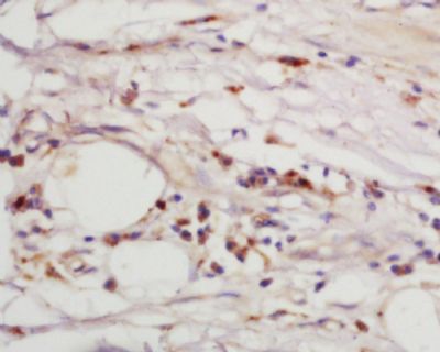
Epsin 1 Polyclonal Antibody
Purified Rabbit Polyclonal Antibody (Pab)
- SPECIFICATION
- CITATIONS
- PROTOCOLS
- BACKGROUND

Application
| WB, IHC-P, IHC-F, IF, ICC, E |
|---|---|
| Primary Accession | Q9Y6I3 |
| Reactivity | Rat, Dog |
| Host | Rabbit |
| Clonality | Polyclonal |
| Calculated MW | 60 KDa |
| Physical State | Liquid |
| Immunogen | KLH conjugated synthetic peptide derived from human Epsin 1/EPN1 |
| Epitope Specificity | 101-200/576 |
| Isotype | IgG |
| Purity | affinity purified by Protein A |
| Buffer | 0.01M TBS (pH7.4) with 1% BSA, 0.02% Proclin300 and 50% Glycerol. |
| SUBCELLULAR LOCATION | Cytoplasm (By similarity). Cell membrane; Peripheral membrane protein (By similarity). Nucleus (By similarity). Membrane, clathrin-coated pit (By similarity). Note=Associated with the cytoplasmic membrane at sites where clathrin-coated pits are forming. Colocalizes with clathrin and AP-2 in a punctate pattern on the plasma membrane. Detected in presynaptic nerve terminals and in Golgi stacks. May shuttle to the nucleus when associated with ZBTB16/ZNF145 (By similarity). |
| SIMILARITY | Belongs to the epsin family. Contains 1 ENTH (epsin N-terminal homology) domain. Contains 3 UIM (ubiquitin-interacting motif) repeats. |
| SUBUNIT | Monomer. Binds clathrin, ZBTB16/ZNF145 and ITSN1. Binds ubiquitinated proteins (By similarity). Binds REPS2, EPS15, AP2A1 and AP2A2. Interacts with RALBP1 in a complex also containing NUMB and TFAP2A during interphase and mitosis. Interacts with AP2B1. [SUBCELLULAR LOCATION] |
| Post-translational modifications | Phosphorylated on serine and/or threonine residues in mitotic cells. Phosphorylation reduces interaction with REPS2, AP-2 and the membrane fraction. Depolarization of synaptosomes results in dephosphorylation. Ubiquitinated (By similarity). |
| Important Note | This product as supplied is intended for research use only, not for use in human, therapeutic or diagnostic applications. |
| Background Descriptions | Epsin 1 is an endocytic accessory protein, with significant similarity to the Xenopus mitotic phosphoprotein MP90. Epsin 1 interacts with Eps15 (the ?subunit of the Clathrin adaptor AP2), Clathrin and other accessory proteins. The mitotic phosphorylation of these proteins may be one of the mechanisms by which the invagination of Clathrin-coated pits is blocked in mitosis. Both Epsin and Eps15, like other cytosolic components of the synaptic vesicle endo-cytic machinery, undergo constitutive phosphorylation and depolarization-dependent dephosphorylation in nerve terminals. Epsin 1 also contributes to the mechanism of Clathrin-vesicle-dependent endocytosis. The human Epsin 1 protein contains an Epsin N-terminal homology (ENTH) region and a single Clathrin-binding (LVDLD) motif. Epsin 1 localizes to the leading edge of a vesicular coated pit where the membrane is being actively bent. |
| Gene ID | 29924 |
|---|---|
| Other Names | Epsin-1, EH domain-binding mitotic phosphoprotein, EPS-15-interacting protein 1, EPN1 |
| Dilution | WB=1:500-2000,IHC-P=1:100-500,IHC-F=1:100-500,ICC=1:50,IF=1:100-500,ELISA=1:5000-10000 |
| Storage | Store at -20 ℃ for one year. Avoid repeated freeze/thaw cycles. When reconstituted in sterile pH 7.4 0.01M PBS or diluent of antibody the antibody is stable for at least two weeks at 2-4 ℃. |
| Name | EPN1 |
|---|---|
| Function | Binds to membranes enriched in phosphatidylinositol 4,5- bisphosphate (PtdIns(4,5)P2). Modifies membrane curvature and facilitates the formation of clathrin-coated invaginations (By similarity). Regulates receptor-mediated endocytosis (PubMed:10393179, PubMed:10557078). |
| Cellular Location | Cytoplasm. Cell membrane; Peripheral membrane protein. Nucleus. Membrane, clathrin-coated pit Note=Associated with the cytoplasmic membrane at sites where clathrin- coated pits are forming. Colocalizes with clathrin and AP-2 in a punctate pattern on the plasma membrane. Detected in presynaptic nerve terminals and in Golgi stacks. May shuttle to the nucleus when associated with ZBTB16/ZNF145 (By similarity). |
Research Areas
Citations (0)

Thousands of laboratories across the world have published research that depended on the performance of antibodies from Abcepta to advance their research. Check out links to articles that cite our products in major peer-reviewed journals, organized by research category.
Submit your citation using an Abcepta antibody to
info@abcepta.com, and receive a free "I Love Antibodies" mug.
info@abcepta.com, and receive a free "I Love Antibodies" mug.

Application Protocols
Provided below are standard protocols that you may find useful for product applications.
Abcepta welcomes feedback from its customers.
If you have used an Abcepta product and would like to share how it has performed, please click on the "Submit Review" button and provide the requested information. Our staff will examine and post your review and contact you if needed.
If you have any additional inquiries please email technical services at tech@abcepta.com.
$ 385.00
Cat# AP55059
Ordering Information
United States
AlbaniaAustraliaAustriaBelgiumBosnia & HerzegovinaBrazilBulgariaCanadaCentral AmericaChinaCroatiaCyprusCzech RepublicDenmarkEstoniaFinlandFranceGermanyGreeceHong KongHungaryIcelandIndiaIndonesiaIrelandIsraelItalyJapanLatviaLithuaniaLuxembourgMacedoniaMalaysiaMaltaMexicoNetherlandsNew ZealandNorwayPakistanPolandPortugalRomaniaSerbiaSingaporeSlovakiaSloveniaSouth AfricaSouth KoreaSpainSwedenSwitzerlandTaiwanTurkeyUnited KingdomUnited StatesVietnamWorldwideOthers
USA Headquarters
(888) 735-7227 / (858) 622-0099 or (858) 875-1900
Other Products
Shipping Information
Domestic orders (in stock items)
Shipped out the same day. Orders placed after 1 PM (PST) will ship out the next business day.
International orders
Contact your local distributors
