


 Foundational characteristics of cancer include proliferation, angiogenesis, migration, evasion of apoptosis, and cellular immortality. Find key markers for these cellular processes and antibodies to detect them.
 The SUMOplot™ Analysis Program predicts and scores sumoylation sites in your protein. SUMOylation is a post-translational modification involved in various cellular processes, such as nuclear-cytosolic transport, transcriptional regulation, apoptosis, protein stability, response to stress, and progression through the cell cycle.
 The Autophagy Receptor Motif Plotter predicts and scores autophagy receptor binding sites in your protein. Identifying proteins connected to this pathway is critical to understanding the role of autophagy in physiological as well as pathological processes such as development, differentiation, neurodegenerative diseases, stress, infection, and cancer.
Optineurin Polyclonal Antibody
Purified Rabbit Polyclonal Antibody (Pab)
- SPECIFICATION
- CITATIONS
- PROTOCOLS
- BACKGROUND

Application
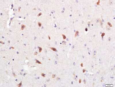
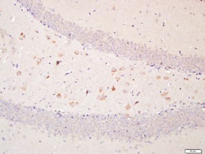
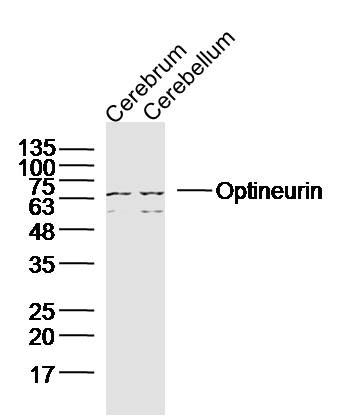
| WB, IHC-P, IHC-F, IF, ICC, E |
|---|---|
| Primary Accession | Q96CV9 |
| Reactivity | Rat, Bovine |
| Host | Rabbit |
| Clonality | Polyclonal |
| Calculated MW | 66 KDa |
| Physical State | Liquid |
| Immunogen | KLH conjugated synthetic peptide derived from human Optineurin |
| Epitope Specificity | 341-440/577 |
| Isotype | IgG |
| Purity | affinity purified by Protein A |
| Buffer | 0.01M TBS (pH7.4) with 1% BSA, 0.02% Proclin300 and 50% Glycerol. |
| SUBCELLULAR LOCATION | Cytoplasm > perinuclear region. Golgi apparatus. Golgi apparatus > trans-Golgi network. Found in the perinuclear region and associates with the Golgi apparatus. Colocalizes with MYO6 and RAB8 at the Golgi complex and in vesicular structures close to the plasma membrane. |
| Post-translational modifications | Phosphorylated. Phosphorylation is induced by phorbol esters and decreases its half-time. |
| DISEASE | Defects in OPTN are the cause of primary open angle glaucoma type 1E (GLC1E) [MIM:137760]. Primary open angle glaucoma (POAG) is characterized by a specific pattern of optic nerve and visual field defects. The angle of the anterior chamber of the eye is open, and usually the intraocular pressure is increased. The disease is asymptomatic until the late stages, by which time significant and irreversible optic nerve damage has already taken place. Defects in OPTN are a cause of susceptibility to normal pressure glaucoma (NPG) [MIM:606657]. Defects in OPTN are the cause of amyotrophic lateral sclerosis type 12 (ALS12) [MIM:613435]. It is a neurodegenerative disorder affecting upper motor neurons in the brain and lower motor neurons in the brain stem and spinal cord, resulting in fatal paralysis. Sensory abnormalities are absent. Death usually occurs within 2 to 5 years. The etiology of amyotrophic lateral sclerosis is likely to be multifactorial, involving both genetic and environmental factors. The disease is inherited in 5-10% of the cases. |
| Important Note | This product as supplied is intended for research use only, not for use in human, therapeutic or diagnostic applications. |
| Background Descriptions | This gene encodes the coiled-coil containing protein optineurin. Optineurin may play a role in normal-tension glaucoma and adult-onset primary open angle glaucoma. Optineurin interacts with adenovirus E3-14.7K protein and may utilize tumor necrosis factor-alpha or Fas-ligand pathways to mediate apoptosis, inflammation or vasoconstriction. Optineurin may also function in cellular morphogenesis and membrane trafficking, vesicle trafficking, and transcription activation through its interactions with the RAB8, huntingtin, and transcription factor IIIA proteins. Alternative splicing results in multiple transcript variants encoding the same protein. [provided by RefSeq, Jul 2008] |
| Gene ID | 10133 |
|---|---|
| Other Names | Optineurin, E3-14.7K-interacting protein, FIP-2, Huntingtin yeast partner L, Huntingtin-interacting protein 7, HIP-7, Huntingtin-interacting protein L, NEMO-related protein, Optic neuropathy-inducing protein, Transcription factor IIIA-interacting protein, TFIIIA-IntP, OPTN, FIP2, GLC1E, HIP7, HYPL, NRP |
| Target/Specificity | Present in acqueous humor of the eye (at protein level). Highly expressed in trabecular meshwork. Expressed nonpigmented ciliary epithelium, retina, brain, adrenal cortex, fetus, lymphocyte, fibroblast, skeletal muscle, heart, liver, brain and placenta. |
| Dilution | WB=1:500-2000,IHC-P=1:100-500,IHC-F=1:100-500,ICC=1:100-500,IF=1:100-500,ELISA=1:5000-10000 |
| Storage | Store at -20 ℃ for one year. Avoid repeated freeze/thaw cycles. When reconstituted in sterile pH 7.4 0.01M PBS or diluent of antibody the antibody is stable for at least two weeks at 2-4 ℃. |
| Name | OPTN |
|---|---|
| Function | Plays an important role in the maintenance of the Golgi complex, in membrane trafficking, in exocytosis, through its interaction with myosin VI and Rab8 (PubMed:27534431). Links myosin VI to the Golgi complex and plays an important role in Golgi ribbon formation (PubMed:27534431). Plays a role in the activation of innate immune response during viral infection. Mechanistically, recruits TBK1 at the Golgi apparatus, promoting its trans-phosphorylation after RLR or TLR3 stimulation (PubMed:27538435). In turn, activated TBK1 phosphorylates its downstream partner IRF3 to produce IFN-beta/IFNB1. Plays a neuroprotective role in the eye and optic nerve. May act by regulating membrane trafficking and cellular morphogenesis via a complex that contains Rab8 and huntingtin (HD). Mediates the interaction of Rab8 with the probable GTPase-activating protein TBC1D17 during Rab8-mediated endocytic trafficking, such as that of transferrin receptor (TFRC/TfR); regulates Rab8 recruitment to tubules emanating from the endocytic recycling compartment (PubMed:22854040). Autophagy receptor that interacts directly with both the cargo to become degraded and an autophagy modifier of the MAP1 LC3 family; targets ubiquitin- coated bacteria (xenophagy), such as cytoplasmic Salmonella enterica, and appears to function in the same pathway as SQSTM1 and CALCOCO2/NDP52. |
| Cellular Location | Cytoplasm, perinuclear region. Golgi apparatus. Golgi apparatus, trans-Golgi network Cytoplasmic vesicle, autophagosome. Cytoplasmic vesicle. Recycling endosome. Note=Found in the perinuclear region and associates with the Golgi apparatus (PubMed:27534431) Colocalizes with MYO6 and RAB8 at the Golgi complex and in vesicular structures close to the plasma membrane. Localizes to LC3-positive cytoplasmic vesicles upon induction of autophagy |
| Tissue Location | Present in aqueous humor of the eye (at protein level). Expressed in the trabecular meshwork (at protein level) (PubMed:11834836, PubMed:12379221, PubMed:12646749). Expressed in nonpigmented ciliary epithelium (at protein level) (PubMed:11834836) Expressed at high levels in skeletal muscle, also detected in heart, brain, pancreas, kidney, placenta and liver (PubMed:9488477). Expressed in dermal fibroblasts (at protein level) (PubMed:11834836) |
Research Areas
Citations (0)

Thousands of laboratories across the world have published research that depended on the performance of antibodies from Abcepta to advance their research. Check out links to articles that cite our products in major peer-reviewed journals, organized by research category.
Submit your citation using an Abcepta antibody to
info@abcepta.com, and receive a free "I Love Antibodies" mug.
info@abcepta.com, and receive a free "I Love Antibodies" mug.

Application Protocols
Provided below are standard protocols that you may find useful for product applications.
Abcepta welcomes feedback from its customers.
If you have used an Abcepta product and would like to share how it has performed, please click on the "Submit Review" button and provide the requested information. Our staff will examine and post your review and contact you if needed.
If you have any additional inquiries please email technical services at tech@abcepta.com.
$ 385.00
Cat# AP55234
Ordering Information
United States
AlbaniaAustraliaAustriaBelgiumBosnia & HerzegovinaBrazilBulgariaCanadaCentral AmericaChinaCroatiaCyprusCzech RepublicDenmarkEstoniaFinlandFranceGermanyGreeceHong KongHungaryIcelandIndiaIndonesiaIrelandIsraelItalyJapanLatviaLithuaniaLuxembourgMacedoniaMalaysiaMaltaMexicoNetherlandsNew ZealandNorwayPakistanPolandPortugalRomaniaSerbiaSingaporeSlovakiaSloveniaSouth AfricaSouth KoreaSpainSwedenSwitzerlandTaiwanTurkeyUnited KingdomUnited StatesVietnamWorldwideOthers
USA Headquarters
(888) 735-7227 / (858) 622-0099 or (858) 875-1900
Other Products
Shipping Information
Domestic orders (in stock items)
Shipped out the same day. Orders placed after 1 PM (PST) will ship out the next business day.
International orders
Contact your local distributors
