


 Foundational characteristics of cancer include proliferation, angiogenesis, migration, evasion of apoptosis, and cellular immortality. Find key markers for these cellular processes and antibodies to detect them.
 The SUMOplot™ Analysis Program predicts and scores sumoylation sites in your protein. SUMOylation is a post-translational modification involved in various cellular processes, such as nuclear-cytosolic transport, transcriptional regulation, apoptosis, protein stability, response to stress, and progression through the cell cycle.
 The Autophagy Receptor Motif Plotter predicts and scores autophagy receptor binding sites in your protein. Identifying proteins connected to this pathway is critical to understanding the role of autophagy in physiological as well as pathological processes such as development, differentiation, neurodegenerative diseases, stress, infection, and cancer.
HGD Polyclonal Antibody
Purified Rabbit Polyclonal Antibody (Pab)
- SPECIFICATION
- CITATIONS
- PROTOCOLS
- BACKGROUND

Application
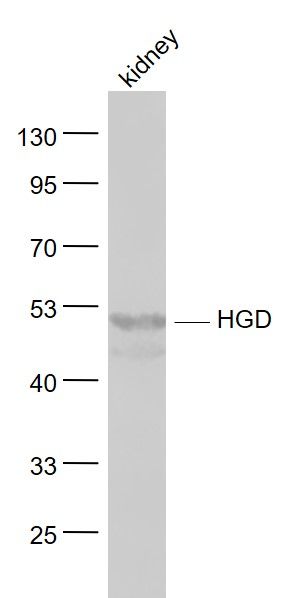
| WB, IHC-P, IHC-F, IF, ICC, E |
|---|---|
| Primary Accession | Q93099 |
| Reactivity | Rat |
| Host | Rabbit |
| Clonality | Polyclonal |
| Calculated MW | 50 KDa |
| Physical State | Liquid |
| Immunogen | KLH conjugated synthetic peptide derived from human HGD |
| Epitope Specificity | 351-445/445 |
| Isotype | IgG |
| Purity | affinity purified by Protein A |
| Buffer | 0.01M TBS (pH7.4) with 1% BSA, 0.02% Proclin300 and 50% Glycerol. |
| SIMILARITY | Belongs to the homogentisate dioxygenase family. |
| DISEASE | Alkaptonuria (AKU) [MIM:203500]: An autosomal recessive error of metabolism characterized by an increase in the level of homogentisic acid. The clinical manifestations are urine that turns dark on standing and alkalinization, black ochronotic pigmentation of cartilage and collagenous tissues, and spine arthritis. Note=The disease is caused by mutations affecting the gene represented in this entry. |
| Important Note | This product as supplied is intended for research use only, not for use in human, therapeutic or diagnostic applications. |
| Background Descriptions | HGD is a 445 amino acid protein that belongs to the homogentisate dioxygenase family and is involved in the pathway of amino acid degradation. Expressed at high levels in kidney, colon, liver, prostate and small intestine, HGD uses iron as a cofactor to catalyze the oxygen-dependent conversion of homogentisate to 4-maleylacetoacetate, a reaction that is the fourth step in the creation of L-phenylalanine from fumarate and acetoacetic acid. Defects in the gene encoding HGD are the cause of alkaptonuria (AKU), an autosomal recessive disorder that is characterized by urine that turns dark on standing and alkalinization, black ochronotic pigmentation of cartilage and collagenous tissues and spine arthritis. |
| Gene ID | 3081 |
|---|---|
| Other Names | Homogentisate 1, 2-dioxygenase, 1.13.11.5, Homogentisate oxygenase, Homogentisic acid oxidase, Homogentisicase, HGD, HGO |
| Target/Specificity | Highest expression in the prostate, small intestine, colon, kidney and liver. |
| Dilution | WB=1:500-2000,IHC-P=1:100-500,IHC-F=1:100-500,ICC=1:100-500,IF=1:100-500,ELISA=1:5000-10000 |
| Storage | Store at -20 ℃ for one year. Avoid repeated freeze/thaw cycles. When reconstituted in sterile pH 7.4 0.01M PBS or diluent of antibody the antibody is stable for at least two weeks at 2-4 ℃. |
| Name | HGD |
|---|---|
| Synonyms | HGO |
| Function | Catalyzes the conversion of homogentisate to maleylacetoacetate. |
| Tissue Location | Highest expression in the prostate, small intestine, colon, kidney and liver |
Research Areas
Citations (0)

Thousands of laboratories across the world have published research that depended on the performance of antibodies from Abcepta to advance their research. Check out links to articles that cite our products in major peer-reviewed journals, organized by research category.
Submit your citation using an Abcepta antibody to
info@abcepta.com, and receive a free "I Love Antibodies" mug.
info@abcepta.com, and receive a free "I Love Antibodies" mug.

Application Protocols
Provided below are standard protocols that you may find useful for product applications.
Abcepta welcomes feedback from its customers.
If you have used an Abcepta product and would like to share how it has performed, please click on the "Submit Review" button and provide the requested information. Our staff will examine and post your review and contact you if needed.
If you have any additional inquiries please email technical services at tech@abcepta.com.
$ 385.00
Cat# AP56013
Ordering Information
United States
AlbaniaAustraliaAustriaBelgiumBosnia & HerzegovinaBrazilBulgariaCanadaCentral AmericaChinaCroatiaCyprusCzech RepublicDenmarkEstoniaFinlandFranceGermanyGreeceHong KongHungaryIcelandIndiaIndonesiaIrelandIsraelItalyJapanLatviaLithuaniaLuxembourgMacedoniaMalaysiaMaltaMexicoNetherlandsNew ZealandNorwayPakistanPolandPortugalRomaniaSerbiaSingaporeSlovakiaSloveniaSouth AfricaSouth KoreaSpainSwedenSwitzerlandTaiwanTurkeyUnited KingdomUnited StatesVietnamWorldwideOthers
USA Headquarters
(888) 735-7227 / (858) 622-0099 or (858) 875-1900
Other Products
Shipping Information
Domestic orders (in stock items)
Shipped out the same day. Orders placed after 1 PM (PST) will ship out the next business day.
International orders
Contact your local distributors
