


 Foundational characteristics of cancer include proliferation, angiogenesis, migration, evasion of apoptosis, and cellular immortality. Find key markers for these cellular processes and antibodies to detect them.
 The SUMOplot™ Analysis Program predicts and scores sumoylation sites in your protein. SUMOylation is a post-translational modification involved in various cellular processes, such as nuclear-cytosolic transport, transcriptional regulation, apoptosis, protein stability, response to stress, and progression through the cell cycle.
 The Autophagy Receptor Motif Plotter predicts and scores autophagy receptor binding sites in your protein. Identifying proteins connected to this pathway is critical to understanding the role of autophagy in physiological as well as pathological processes such as development, differentiation, neurodegenerative diseases, stress, infection, and cancer.
> home > Products > Primary Antibodies > Signal Transduction > Granuphilin/SYTL4 Polyclonal Antibody
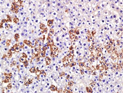
Granuphilin/SYTL4 Polyclonal Antibody
Purified Rabbit Polyclonal Antibody (Pab)
- SPECIFICATION
- CITATIONS
- PROTOCOLS
- BACKGROUND

Application
| IHC-P, IHC-F, IF, ICC, E |
|---|---|
| Primary Accession | Q96C24 |
| Reactivity | Rat, Dog, Bovine |
| Host | Rabbit |
| Clonality | Polyclonal |
| Calculated MW | 76 KDa |
| Physical State | Liquid |
| Immunogen | KLH conjugated synthetic peptide derived from human Granuphilin/SYTL4 |
| Epitope Specificity | 25-125/671 |
| Isotype | IgG |
| Purity | affinity purified by Protein A |
| Buffer | 0.01M TBS (pH7.4) with 1% BSA, 0.02% Proclin300 and 50% Glycerol. |
| SUBCELLULAR LOCATION | Membrane. Cell membrane. Cytoplasmic vesicle > secretory vesicle membrane. Detected close to the plasma membrane and on secretory granules. In pancreas, interacts with insulin-containing vesicles. |
| SIMILARITY | Contains 2 C2 domains. Contains 1 FYVE-type zinc finger. Contains 1 RabBD (Rab-binding) domain. |
| SUBUNIT | Part of a ternary complex containing STX1A and RAB27A. Can bind both dominant negative and dominant active mutants of RAB27A. Binds STXBP1, RAB3A, RAB8A and RAB27B. Interacts with MYO5A (By similarity). |
| Important Note | This product as supplied is intended for research use only, not for use in human, therapeutic or diagnostic applications. |
| Background Descriptions | This gene encodes a member of the synaptotagmin like protein family. Members of this family are characterized by an N-terminal Rab27 binding domain and C-terminal tandem C2 domains. The encoded protein binds specific small Rab GTPases and is involved in intracellular membrane trafficking. This protein binds Rab27 and may be involved in inhibiting dense core vesicle exocytosis. Alternate splicing results in multiple transcript variants that encode the same protein. [provided by RefSeq, Mar 2010] |
| Gene ID | 94121 |
|---|---|
| Other Names | Synaptotagmin-like protein 4, Exophilin-2, Granuphilin, SYTL4 |
| Dilution | IHC-P=1:100-500,IHC-F=1:100-500,ICC=1:100-500,IF=1:100-500,ELISA=1:5000-10000 |
| Storage | Store at -20 ℃ for one year. Avoid repeated freeze/thaw cycles. When reconstituted in sterile pH 7.4 0.01M PBS or diluent of antibody the antibody is stable for at least two weeks at 2-4 ℃. |
| Name | SYTL4 |
|---|---|
| Function | Modulates exocytosis of dense-core granules and secretion of hormones in the pancreas and the pituitary. Interacts with vesicles containing negatively charged phospholipids in a Ca(2+)-independent manner (By similarity). |
| Cellular Location | Membrane; Peripheral membrane protein. Cell membrane; Peripheral membrane protein. Cytoplasmic vesicle, secretory vesicle membrane; Peripheral membrane protein. Note=Detected close to the plasma membrane and on secretory granules. In pancreas, interacts with insulin-containing vesicles (By similarity) |
Research Areas
Citations (0)

Thousands of laboratories across the world have published research that depended on the performance of antibodies from Abcepta to advance their research. Check out links to articles that cite our products in major peer-reviewed journals, organized by research category.
Submit your citation using an Abcepta antibody to
info@abcepta.com, and receive a free "I Love Antibodies" mug.
info@abcepta.com, and receive a free "I Love Antibodies" mug.

Application Protocols
Provided below are standard protocols that you may find useful for product applications.
Abcepta welcomes feedback from its customers.
If you have used an Abcepta product and would like to share how it has performed, please click on the "Submit Review" button and provide the requested information. Our staff will examine and post your review and contact you if needed.
If you have any additional inquiries please email technical services at tech@abcepta.com.
$ 385.00
Cat# AP56214
Ordering Information
United States
AlbaniaAustraliaAustriaBelgiumBosnia & HerzegovinaBrazilBulgariaCanadaCentral AmericaChinaCroatiaCyprusCzech RepublicDenmarkEstoniaFinlandFranceGermanyGreeceHong KongHungaryIcelandIndiaIndonesiaIrelandIsraelItalyJapanLatviaLithuaniaLuxembourgMacedoniaMalaysiaMaltaMexicoNetherlandsNew ZealandNorwayPakistanPolandPortugalRomaniaSerbiaSingaporeSlovakiaSloveniaSouth AfricaSouth KoreaSpainSwedenSwitzerlandTaiwanTurkeyUnited KingdomUnited StatesVietnamWorldwideOthers
USA Headquarters
(888) 735-7227 / (858) 622-0099 or (858) 875-1900
Other Products
Shipping Information
Domestic orders (in stock items)
Shipped out the same day. Orders placed after 1 PM (PST) will ship out the next business day.
International orders
Contact your local distributors
