


 Foundational characteristics of cancer include proliferation, angiogenesis, migration, evasion of apoptosis, and cellular immortality. Find key markers for these cellular processes and antibodies to detect them.
 The SUMOplot™ Analysis Program predicts and scores sumoylation sites in your protein. SUMOylation is a post-translational modification involved in various cellular processes, such as nuclear-cytosolic transport, transcriptional regulation, apoptosis, protein stability, response to stress, and progression through the cell cycle.
 The Autophagy Receptor Motif Plotter predicts and scores autophagy receptor binding sites in your protein. Identifying proteins connected to this pathway is critical to understanding the role of autophagy in physiological as well as pathological processes such as development, differentiation, neurodegenerative diseases, stress, infection, and cancer.
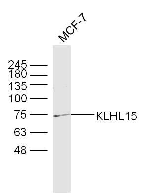
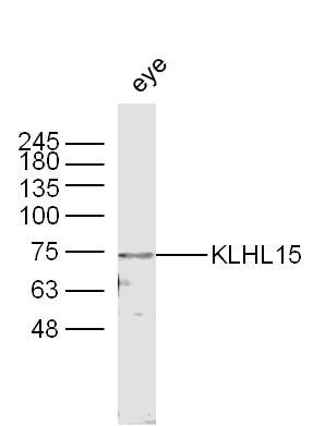
KLHL15 Polyclonal Antibody
Purified Rabbit Polyclonal Antibody (Pab)
- SPECIFICATION
- CITATIONS
- PROTOCOLS
- BACKGROUND

Application
| WB, IHC-P, IHC-F, IF, ICC, E |
|---|---|
| Primary Accession | Q96M94 |
| Reactivity | Rat, Pig, Bovine |
| Host | Rabbit |
| Clonality | Polyclonal |
| Calculated MW | 70 KDa |
| Physical State | Liquid |
| Immunogen | KLH conjugated synthetic peptide derived from human KLHL15 |
| Epitope Specificity | 401-500/604 |
| Isotype | IgG |
| Purity | affinity purified by Protein A |
| Buffer | 0.01M TBS (pH7.4) with 1% BSA, 0.02% Proclin300 and 50% Glycerol. |
| SIMILARITY | Contains 1 BACK (BTB/Kelch associated) domain. Contains 1 BTB (POZ) domain. Contains 5 Kelch repeats. |
| SUBUNIT | Interacts with CUL3. |
| Important Note | This product as supplied is intended for research use only, not for use in human, therapeutic or diagnostic applications. |
| Background Descriptions | KLHL15 is believed to be a substrate-specific adapter of an E3 ubiquitin-protein ligase complex which regulates the ubiquitination, and subsequent proteasomal degradation, of target proteins. KLHL15 contains one BACK (BTB/Kelch associated) domain, five kelch repeats and one BTB domain. The BTB (broad complex, tramtrack and bric-a-brac) domain, also known as the POZ (poxvirus and zinc finger) domain, is an N-terminal homodimerization domain that contains multiple copies of kelch repeats and/or C2H2-type zinc fingers. Proteins that contain BTB domains are thought to be involved in transcriptional regulation via control of chromatin structure and function. |
| Gene ID | 80311 |
|---|---|
| Other Names | Kelch-like protein 15, KLHL15, KIAA1677 |
| Dilution | WB=1:500-2000,IHC-P=1:100-500,IHC-F=1:100-500,ICC=1:100-500,IF=1:100-500,ELISA=1:5000-10000 |
| Storage | Store at -20 ℃ for one year. Avoid repeated freeze/thaw cycles. When reconstituted in sterile pH 7.4 0.01M PBS or diluent of antibody the antibody is stable for at least two weeks at 2-4 ℃. |
| Name | KLHL15 |
|---|---|
| Synonyms | KIAA1677 |
| Function | Substrate-specific adapter for CUL3 E3 ubiquitin-protein ligase complex (PubMed:14528312, PubMed:27561354, PubMed:35219381). Acts as an adapter for CUL3 to target the serine/threonine-protein phosphatase 2A (PP2A) subunit PPP2R5B for ubiquitination and subsequent proteasomal degradation, thus promoting exchange with other regulatory subunits (PubMed:23135275). Acts as an adapter for CUL3 to target the DNA-end resection factor RBBP8/CtIP for ubiquitination and subsequent proteasomal degradation (PubMed:27561354, PubMed:35219381). Through the regulation of RBBP8/CtIP protein turnover, plays a key role in DNA damage response, favoring DNA double-strand repair through error-prone non-homologous end joining (NHEJ) over error-free, RBBP8-mediated homologous recombination (HR) (PubMed:27561354, PubMed:35219381). |
| Cellular Location | Nucleus. |
Research Areas
Citations (0)

Thousands of laboratories across the world have published research that depended on the performance of antibodies from Abcepta to advance their research. Check out links to articles that cite our products in major peer-reviewed journals, organized by research category.
Submit your citation using an Abcepta antibody to
info@abcepta.com, and receive a free "I Love Antibodies" mug.
info@abcepta.com, and receive a free "I Love Antibodies" mug.

Application Protocols
Provided below are standard protocols that you may find useful for product applications.
Abcepta welcomes feedback from its customers.
If you have used an Abcepta product and would like to share how it has performed, please click on the "Submit Review" button and provide the requested information. Our staff will examine and post your review and contact you if needed.
If you have any additional inquiries please email technical services at tech@abcepta.com.
$ 385.00
Cat# AP56394
Ordering Information
United States
AlbaniaAustraliaAustriaBelgiumBosnia & HerzegovinaBrazilBulgariaCanadaCentral AmericaChinaCroatiaCyprusCzech RepublicDenmarkEstoniaFinlandFranceGermanyGreeceHong KongHungaryIcelandIndiaIndonesiaIrelandIsraelItalyJapanLatviaLithuaniaLuxembourgMacedoniaMalaysiaMaltaMexicoNetherlandsNew ZealandNorwayPakistanPolandPortugalRomaniaSerbiaSingaporeSlovakiaSloveniaSouth AfricaSouth KoreaSpainSwedenSwitzerlandTaiwanTurkeyUnited KingdomUnited StatesVietnamWorldwideOthers
USA Headquarters
(888) 735-7227 / (858) 622-0099 or (858) 875-1900
Other Products
Shipping Information
Domestic orders (in stock items)
Shipped out the same day. Orders placed after 1 PM (PST) will ship out the next business day.
International orders
Contact your local distributors
