


 Foundational characteristics of cancer include proliferation, angiogenesis, migration, evasion of apoptosis, and cellular immortality. Find key markers for these cellular processes and antibodies to detect them.
 The SUMOplot™ Analysis Program predicts and scores sumoylation sites in your protein. SUMOylation is a post-translational modification involved in various cellular processes, such as nuclear-cytosolic transport, transcriptional regulation, apoptosis, protein stability, response to stress, and progression through the cell cycle.
 The Autophagy Receptor Motif Plotter predicts and scores autophagy receptor binding sites in your protein. Identifying proteins connected to this pathway is critical to understanding the role of autophagy in physiological as well as pathological processes such as development, differentiation, neurodegenerative diseases, stress, infection, and cancer.
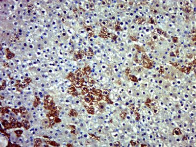
HKDC1 Polyclonal Antibody
Purified Rabbit Polyclonal Antibody (Pab)
- SPECIFICATION
- CITATIONS
- PROTOCOLS
- BACKGROUND

Application
| IHC-P, IHC-F, IF, ICC |
|---|---|
| Primary Accession | Q2TB90 |
| Reactivity | Rat, Pig, Dog, Bovine |
| Host | Rabbit |
| Clonality | Polyclonal |
| Calculated MW | 103 KDa |
| Physical State | Liquid |
| Immunogen | KLH conjugated synthetic peptide derived from human HKDC1 |
| Epitope Specificity | 21-120/917 |
| Isotype | IgG |
| Purity | affinity purified by Protein A |
| Buffer | 0.01M TBS (pH7.4) with 1% BSA, 0.02% Proclin300 and 50% Glycerol. |
| SIMILARITY | Belongs to the hexokinase family. |
| Important Note | This product as supplied is intended for research use only, not for use in human, therapeutic or diagnostic applications. |
| Gene ID | 80201 |
|---|---|
| Other Names | Hexokinase HKDC1, 2.7.1.1, Hexokinase domain-containing protein 1, HKDC1 (HGNC:23302) |
| Dilution | IHC-P=1:100-500,IHC-F=1:100-500,ICC=1:100-500,IF=1:100-500 |
| Storage | Store at -20 ℃ for one year. Avoid repeated freeze/thaw cycles. When reconstituted in sterile pH 7.4 0.01M PBS or diluent of antibody the antibody is stable for at least two weeks at 2-4 ℃. |
| Name | HKDC1 (HGNC:23302) |
|---|---|
| Function | Catalyzes the phosphorylation of hexose to hexose 6- phosphate, although at very low level compared to other hexokinases (PubMed:30517626). Has low glucose phosphorylating activity compared to other hexokinases (PubMed:30517626). Involved in glucose homeostasis and hepatic lipid accumulation. Required to maintain whole-body glucose homeostasis during pregnancy; however additional evidences are required to confirm this role (By similarity). |
| Cellular Location | Cytoplasm. Mitochondrion membrane; Peripheral membrane protein. Photoreceptor inner segment {ECO:0000250|UniProtKB:Q91W97}. Note=The mitochondrial-binding peptide (MBP) region promotes association with the mitochondrion |
| Tissue Location | Widely expressed (PubMed:27459389, PubMed:29401404). Highly expressed in the brush border, surface epithelium and the myenteric plexus of the small and large intestines; the acinar centrocytes and interlobular ducts of the pancreas; and the alveolar macrophages in the lungs (at protein level) (PubMed:29401404) Present at moderate level in the thyroid follicular epithelium (at protein level) (PubMed:29401404). |
Research Areas
Citations (0)

Thousands of laboratories across the world have published research that depended on the performance of antibodies from Abcepta to advance their research. Check out links to articles that cite our products in major peer-reviewed journals, organized by research category.
Submit your citation using an Abcepta antibody to
info@abcepta.com, and receive a free "I Love Antibodies" mug.
info@abcepta.com, and receive a free "I Love Antibodies" mug.

Application Protocols
Provided below are standard protocols that you may find useful for product applications.
Abcepta welcomes feedback from its customers.
If you have used an Abcepta product and would like to share how it has performed, please click on the "Submit Review" button and provide the requested information. Our staff will examine and post your review and contact you if needed.
If you have any additional inquiries please email technical services at tech@abcepta.com.
$ 385.00
Cat# AP56703
Ordering Information
United States
AlbaniaAustraliaAustriaBelgiumBosnia & HerzegovinaBrazilBulgariaCanadaCentral AmericaChinaCroatiaCyprusCzech RepublicDenmarkEstoniaFinlandFranceGermanyGreeceHong KongHungaryIcelandIndiaIndonesiaIrelandIsraelItalyJapanLatviaLithuaniaLuxembourgMacedoniaMalaysiaMaltaMexicoNetherlandsNew ZealandNorwayPakistanPolandPortugalRomaniaSerbiaSingaporeSlovakiaSloveniaSouth AfricaSouth KoreaSpainSwedenSwitzerlandTaiwanTurkeyUnited KingdomUnited StatesVietnamWorldwideOthers
USA Headquarters
(888) 735-7227 / (858) 622-0099 or (858) 875-1900
Other Products
Shipping Information
Domestic orders (in stock items)
Shipped out the same day. Orders placed after 1 PM (PST) will ship out the next business day.
International orders
Contact your local distributors
