


 Foundational characteristics of cancer include proliferation, angiogenesis, migration, evasion of apoptosis, and cellular immortality. Find key markers for these cellular processes and antibodies to detect them.
 The SUMOplot™ Analysis Program predicts and scores sumoylation sites in your protein. SUMOylation is a post-translational modification involved in various cellular processes, such as nuclear-cytosolic transport, transcriptional regulation, apoptosis, protein stability, response to stress, and progression through the cell cycle.
 The Autophagy Receptor Motif Plotter predicts and scores autophagy receptor binding sites in your protein. Identifying proteins connected to this pathway is critical to understanding the role of autophagy in physiological as well as pathological processes such as development, differentiation, neurodegenerative diseases, stress, infection, and cancer.
LMBRD1 Polyclonal Antibody
Purified Rabbit Polyclonal Antibody (Pab)
- SPECIFICATION
- CITATIONS
- PROTOCOLS
- BACKGROUND

Application
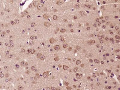
| IHC-P, IHC-F, IF, ICC |
|---|---|
| Primary Accession | Q9NUN5 |
| Reactivity | Rat, Pig, Dog, Bovine |
| Host | Rabbit |
| Clonality | Polyclonal |
| Calculated MW | 61 KDa |
| Physical State | Liquid |
| Immunogen | KLH conjugated synthetic peptide derived from human LMBRD1 |
| Epitope Specificity | 21-120/540 |
| Isotype | IgG |
| Purity | affinity purified by Protein A |
| Buffer | 0.01M TBS (pH7.4) with 1% BSA, 0.02% Proclin300 and 50% Glycerol. |
| SUBCELLULAR LOCATION | Lysosome membrane. |
| SIMILARITY | Belongs to the LIMR family. LMBRD1 subfamily. |
| Post-translational modifications | N-glycosylated. |
| DISEASE | Defects in LMBRD1 are the cause of methylmalonic aciduria and homocystinuria type cblF (MMAFHC) [MIM:277380]; also known as homocystinuria-megaloblastic anemia complementation type F. MMAFHC is a disorder of cobalamin metabolism characterized by decreased levels of the coenzymes adenosylcobalamin (AdoCbl) and methylcobalamin (MeCbl). It is due to accumulation of free cobalamin in lysosomes, thus hindering its conversion to cofactors. Clinical features include developmental delay, stomatitis, glossitis, seizures and methylmalonic aciduria responsive to vitamin B12. |
| Important Note | This product as supplied is intended for research use only, not for use in human, therapeutic or diagnostic applications. |
| Background Descriptions | This gene encodes a lysosomal membrane protein that may be involved in the transport and metabolism of cobalamin. This protein also interacts with the large form of the hepatitis delta antigen and may be required for the nucleocytoplasmic shuttling of the hepatitis delta virus. Mutations in this gene are associated with the vitamin B12 metabolism disorder termed, homocystinuria-megaloblastic anemia complementation type F.[provided by RefSeq, Oct 2009] |
| Gene ID | 55788 |
|---|---|
| Other Names | Lysosomal cobalamin transport escort protein LMBD1, LMBD1, HDAg-L-interacting protein NESI, LMBR1 domain-containing protein 1, Nuclear export signal-interacting protein, LMBRD1 (HGNC:23038), C6orf209, NESI |
| Target/Specificity | Isoform 3 is expressed in liver. |
| Dilution | IHC-P=1:100-500,IHC-F=1:100-500,ICC=1:100-500,IF=1:100-500 |
| Storage | Store at -20 ℃ for one year. Avoid repeated freeze/thaw cycles. When reconstituted in sterile pH 7.4 0.01M PBS or diluent of antibody the antibody is stable for at least two weeks at 2-4 ℃. |
| Name | LMBRD1 (HGNC:23038) |
|---|---|
| Synonyms | C6orf209, NESI |
| Function | Lysosomal membrane chaperone required to export cobalamin (vitamin B12) from the lysosome to the cytosol, allowing its conversion to cofactors (PubMed:19136951). Targets ABCD4 transporter from the endoplasmic reticulum to the lysosome (PubMed:27456980). Then forms a complex with lysosomal ABCD4 and cytoplasmic MMACHC to transport cobalamin across the lysosomal membrane (PubMed:25535791). Acts as an adapter protein which plays an important role in mediating and regulating the internalization of the insulin receptor (INSR) (By similarity). Involved in clathrin-mediated endocytosis of INSR via its interaction with adapter protein complex 2 (By similarity). Essential for the initiation of gastrulation and early formation of mesoderm structures during embryogenesis (By similarity). |
| Cellular Location | Endoplasmic reticulum membrane. Lysosome membrane; Multi-pass membrane protein. Cell membrane {ECO:0000250|UniProtKB:Q8K0B2}; Multi-pass membrane protein. Cytoplasmic vesicle, clathrin-coated vesicle {ECO:0000250|UniProtKB:Q8K0B2} |
| Tissue Location | Isoform 3 is expressed in liver. |
Research Areas
Citations (0)

Thousands of laboratories across the world have published research that depended on the performance of antibodies from Abcepta to advance their research. Check out links to articles that cite our products in major peer-reviewed journals, organized by research category.
Submit your citation using an Abcepta antibody to
info@abcepta.com, and receive a free "I Love Antibodies" mug.
info@abcepta.com, and receive a free "I Love Antibodies" mug.

Application Protocols
Provided below are standard protocols that you may find useful for product applications.
Abcepta welcomes feedback from its customers.
If you have used an Abcepta product and would like to share how it has performed, please click on the "Submit Review" button and provide the requested information. Our staff will examine and post your review and contact you if needed.
If you have any additional inquiries please email technical services at tech@abcepta.com.
$ 385.00
Cat# AP57032
Ordering Information
United States
AlbaniaAustraliaAustriaBelgiumBosnia & HerzegovinaBrazilBulgariaCanadaCentral AmericaChinaCroatiaCyprusCzech RepublicDenmarkEstoniaFinlandFranceGermanyGreeceHong KongHungaryIcelandIndiaIndonesiaIrelandIsraelItalyJapanLatviaLithuaniaLuxembourgMacedoniaMalaysiaMaltaMexicoNetherlandsNew ZealandNorwayPakistanPolandPortugalRomaniaSerbiaSingaporeSlovakiaSloveniaSouth AfricaSouth KoreaSpainSwedenSwitzerlandTaiwanTurkeyUnited KingdomUnited StatesVietnamWorldwideOthers
USA Headquarters
(888) 735-7227 / (858) 622-0099 or (858) 875-1900
Other Products
Shipping Information
Domestic orders (in stock items)
Shipped out the same day. Orders placed after 1 PM (PST) will ship out the next business day.
International orders
Contact your local distributors
