


 Foundational characteristics of cancer include proliferation, angiogenesis, migration, evasion of apoptosis, and cellular immortality. Find key markers for these cellular processes and antibodies to detect them.
 The SUMOplot™ Analysis Program predicts and scores sumoylation sites in your protein. SUMOylation is a post-translational modification involved in various cellular processes, such as nuclear-cytosolic transport, transcriptional regulation, apoptosis, protein stability, response to stress, and progression through the cell cycle.
 The Autophagy Receptor Motif Plotter predicts and scores autophagy receptor binding sites in your protein. Identifying proteins connected to this pathway is critical to understanding the role of autophagy in physiological as well as pathological processes such as development, differentiation, neurodegenerative diseases, stress, infection, and cancer.
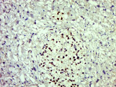
Myt1 Polyclonal Antibody
Purified Rabbit Polyclonal Antibody (Pab)
- SPECIFICATION
- CITATIONS
- PROTOCOLS
- BACKGROUND

Application
| WB, IHC-P, IHC-F, IF, E |
|---|---|
| Primary Accession | Q01538 |
| Reactivity | Rat, Dog, Bovine |
| Host | Rabbit |
| Clonality | Polyclonal |
| Calculated MW | 122 KDa |
| Physical State | Liquid |
| Immunogen | KLH conjugated synthetic peptide derived from human Myt1 |
| Epitope Specificity | 541-640/1121 |
| Isotype | IgG |
| Purity | affinity purified by Protein A |
| Buffer | 0.01M TBS (pH7.4) with 1% BSA, 0.02% Proclin300 and 50% Glycerol. |
| SUBCELLULAR LOCATION | Nucleus. |
| SIMILARITY | Contains 7 C2HC-type zinc fingers. |
| SUBUNIT | nteracts with STEAP3. |
| Important Note | This product as supplied is intended for research use only, not for use in human, therapeutic or diagnostic applications. |
| Background Descriptions | Myt1 is a zinc finger protein that is known to interact with the co-repressor Sin3B and also HDAC1 and HDAC2. The Myt1 family, including Myt1 and Myt1L, exemplifies a class of neural sequence specific transcription factors that actively recruit HDACs to selected genes during CNS development. |
| Gene ID | 4661 |
|---|---|
| Other Names | Myelin transcription factor 1, MyT1, Myelin transcription factor I, MyTI, PLPB1, Proteolipid protein-binding protein, MYT1, KIAA0835, KIAA1050, MTF1, MYTI, PLPB1 |
| Target/Specificity | Mostly in developing nervous system. Expressed in neural progenitors and oligodendrocyte lineage cells. More highly expressed in oligodendrocyte progenitors than in differentiated oligodendrocytes. |
| Dilution | WB=1:500-2000,IHC-P=1:100-500,IHC-F=1:100-500,IF=1:100-500,ELISA=1:5000-10000 |
| Storage | Store at -20 ℃ for one year. Avoid repeated freeze/thaw cycles. When reconstituted in sterile pH 7.4 0.01M PBS or diluent of antibody the antibody is stable for at least two weeks at 2-4 ℃. |
| Name | MYT1 |
|---|---|
| Synonyms | KIAA0835, KIAA1050, MTF1, MYTI, PLPB1 |
| Function | Binds to the promoter region of genes encoding proteolipid proteins of the central nervous system. May play a role in the development of neurons and oligodendroglia in the CNS. May regulate a critical transition point in oligodendrocyte lineage development by modulating oligodendrocyte progenitor proliferation relative to terminal differentiation and up-regulation of myelin gene transcription. |
| Cellular Location | Nucleus. |
| Tissue Location | Mostly in developing nervous system. Expressed in neural progenitors and oligodendrocyte lineage cells. More highly expressed in oligodendrocyte progenitors than in differentiated oligodendrocytes. |
Research Areas
Citations (0)

Thousands of laboratories across the world have published research that depended on the performance of antibodies from Abcepta to advance their research. Check out links to articles that cite our products in major peer-reviewed journals, organized by research category.
Submit your citation using an Abcepta antibody to
info@abcepta.com, and receive a free "I Love Antibodies" mug.
info@abcepta.com, and receive a free "I Love Antibodies" mug.

Application Protocols
Provided below are standard protocols that you may find useful for product applications.
Abcepta welcomes feedback from its customers.
If you have used an Abcepta product and would like to share how it has performed, please click on the "Submit Review" button and provide the requested information. Our staff will examine and post your review and contact you if needed.
If you have any additional inquiries please email technical services at tech@abcepta.com.
$ 385.00
Cat# AP58072
Ordering Information
United States
AlbaniaAustraliaAustriaBelgiumBosnia & HerzegovinaBrazilBulgariaCanadaCentral AmericaChinaCroatiaCyprusCzech RepublicDenmarkEstoniaFinlandFranceGermanyGreeceHong KongHungaryIcelandIndiaIndonesiaIrelandIsraelItalyJapanLatviaLithuaniaLuxembourgMacedoniaMalaysiaMaltaMexicoNetherlandsNew ZealandNorwayPakistanPolandPortugalRomaniaSerbiaSingaporeSlovakiaSloveniaSouth AfricaSouth KoreaSpainSwedenSwitzerlandTaiwanTurkeyUnited KingdomUnited StatesVietnamWorldwideOthers
USA Headquarters
(888) 735-7227 / (858) 622-0099 or (858) 875-1900
Other Products
Shipping Information
Domestic orders (in stock items)
Shipped out the same day. Orders placed after 1 PM (PST) will ship out the next business day.
International orders
Contact your local distributors
