


 Foundational characteristics of cancer include proliferation, angiogenesis, migration, evasion of apoptosis, and cellular immortality. Find key markers for these cellular processes and antibodies to detect them.
 The SUMOplot™ Analysis Program predicts and scores sumoylation sites in your protein. SUMOylation is a post-translational modification involved in various cellular processes, such as nuclear-cytosolic transport, transcriptional regulation, apoptosis, protein stability, response to stress, and progression through the cell cycle.
 The Autophagy Receptor Motif Plotter predicts and scores autophagy receptor binding sites in your protein. Identifying proteins connected to this pathway is critical to understanding the role of autophagy in physiological as well as pathological processes such as development, differentiation, neurodegenerative diseases, stress, infection, and cancer.
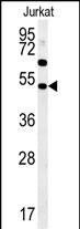
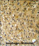
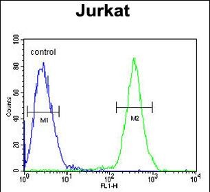
SAMSN1 Antibody (Center)
Affinity Purified Rabbit Polyclonal Antibody (Pab)
- SPECIFICATION
- CITATIONS
- PROTOCOLS
- BACKGROUND

Application
| IHC-P, FC, WB, E |
|---|---|
| Primary Accession | Q9NSI8 |
| Other Accession | NP_071419.3 |
| Reactivity | Human |
| Host | Rabbit |
| Clonality | Polyclonal |
| Isotype | Rabbit IgG |
| Calculated MW | 41708 Da |
| Antigen Region | 255-283 aa |
| Gene ID | 64092 |
|---|---|
| Other Names | SAM domain-containing protein SAMSN-1, Hematopoietic adaptor containing SH3 and SAM domains 1, Nash1, SAM domain, SH3 domain and nuclear localization signals protein 1, SH3-SAM adaptor protein, SAMSN1, HACS1 |
| Target/Specificity | This SAMSN1 antibody is generated from rabbits immunized with a KLH conjugated synthetic peptide between 255-283 amino acids from the Central region of human SAMSN1. |
| Dilution | IHC-P~~1:50~100 FC~~1:10~50 WB~~1:1000 E~~Use at an assay dependent concentration. |
| Format | Purified polyclonal antibody supplied in PBS with 0.09% (W/V) sodium azide. This antibody is purified through a protein A column, followed by peptide affinity purification. |
| Storage | Maintain refrigerated at 2-8°C for up to 2 weeks. For long term storage store at -20°C in small aliquots to prevent freeze-thaw cycles. |
| Precautions | SAMSN1 Antibody (Center) is for research use only and not for use in diagnostic or therapeutic procedures. |
| Name | SAMSN1 |
|---|---|
| Synonyms | HACS1 |
| Function | Negative regulator of B-cell activation. Down-regulates cell proliferation (in vitro). Promotes RAC1-dependent membrane ruffle formation and reorganization of the actin cytoskeleton. Regulates cell spreading and cell polarization. Stimulates HDAC1 activity. Regulates LYN activity by modulating its tyrosine phosphorylation (By similarity). |
| Cellular Location | Nucleus. Cytoplasm. Cell projection, ruffle. Note=Shuttles between cytoplasm and nucleus. Colocalizes with the actin cytoskeleton and actin-rich membrane ruffles (By similarity). |
| Tissue Location | Detected in peripheral blood B-cells (at protein level). Detected in spleen, liver and peripheral blood |
Research Areas
Citations (0)

Thousands of laboratories across the world have published research that depended on the performance of antibodies from Abcepta to advance their research. Check out links to articles that cite our products in major peer-reviewed journals, organized by research category.
Submit your citation using an Abcepta antibody to
info@abcepta.com, and receive a free "I Love Antibodies" mug.
info@abcepta.com, and receive a free "I Love Antibodies" mug.

Application Protocols
Provided below are standard protocols that you may find useful for product applications.
Abcepta welcomes feedback from its customers.
If you have used an Abcepta product and would like to share how it has performed, please click on the "Submit Review" button and provide the requested information. Our staff will examine and post your review and contact you if needed.
If you have any additional inquiries please email technical services at tech@abcepta.com.
$ 150.00
$ 385.00
Cat# AP5877c
Ordering Information
United States
AlbaniaAustraliaAustriaBelgiumBosnia & HerzegovinaBrazilBulgariaCanadaCentral AmericaChinaCroatiaCyprusCzech RepublicDenmarkEstoniaFinlandFranceGermanyGreeceHong KongHungaryIcelandIndiaIndonesiaIrelandIsraelItalyJapanLatviaLithuaniaLuxembourgMacedoniaMalaysiaMaltaMexicoNetherlandsNew ZealandNorwayPakistanPolandPortugalRomaniaSerbiaSingaporeSlovakiaSloveniaSouth AfricaSouth KoreaSpainSwedenSwitzerlandTaiwanTurkeyUnited KingdomUnited StatesVietnamWorldwideOthers
USA Headquarters
(888) 735-7227 / (858) 622-0099 or (858) 875-1900
Other Products
Shipping Information
Domestic orders (in stock items)
Shipped out the same day. Orders placed after 1 PM (PST) will ship out the next business day.
International orders
Contact your local distributors
