


 Foundational characteristics of cancer include proliferation, angiogenesis, migration, evasion of apoptosis, and cellular immortality. Find key markers for these cellular processes and antibodies to detect them.
 The SUMOplot™ Analysis Program predicts and scores sumoylation sites in your protein. SUMOylation is a post-translational modification involved in various cellular processes, such as nuclear-cytosolic transport, transcriptional regulation, apoptosis, protein stability, response to stress, and progression through the cell cycle.
 The Autophagy Receptor Motif Plotter predicts and scores autophagy receptor binding sites in your protein. Identifying proteins connected to this pathway is critical to understanding the role of autophagy in physiological as well as pathological processes such as development, differentiation, neurodegenerative diseases, stress, infection, and cancer.
SAMSN Antibody
- SPECIFICATION
- CITATIONS
- PROTOCOLS
- BACKGROUND

Application
| WB, IF, ICC, E |
|---|---|
| Primary Accession | Q9NSI8 |
| Other Accession | NP_071419, 48762709 |
| Reactivity | Human |
| Host | Rabbit |
| Clonality | Polyclonal |
| Isotype | IgG |
| Calculated MW | 41 kDa |
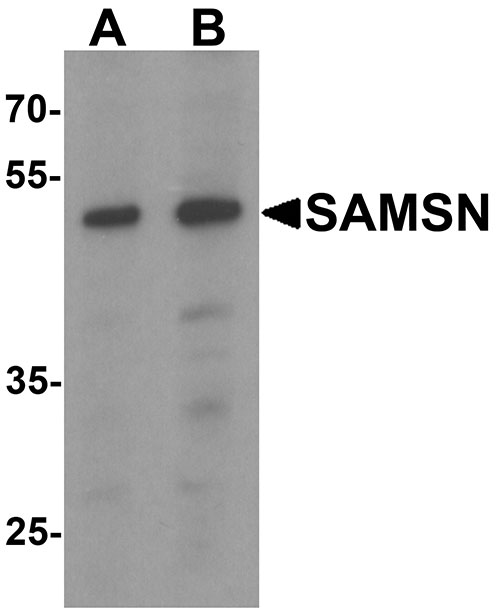
| Application Notes | SAMSN antibody can be used for detection of SAMSN by Western blot at 1 - 2 µg/mL. |
| Gene ID | 64092 |
|---|---|
| Target/Specificity | SAMSN1; At least two isoforms of SAMSN are known to exist. |
| Reconstitution & Storage | SAMSN antibody can be stored at 4℃ for three months and -20℃, stable for up to one year. As with all antibodies care should be taken to avoid repeated freeze thaw cycles. Antibodies should not be exposed to prolonged high temperatures. |
| Precautions | SAMSN Antibody is for research use only and not for use in diagnostic or therapeutic procedures. |
| Name | SAMSN1 |
|---|---|
| Synonyms | HACS1 |
| Function | Negative regulator of B-cell activation. Down-regulates cell proliferation (in vitro). Promotes RAC1-dependent membrane ruffle formation and reorganization of the actin cytoskeleton. Regulates cell spreading and cell polarization. Stimulates HDAC1 activity. Regulates LYN activity by modulating its tyrosine phosphorylation (By similarity). |
| Cellular Location | Nucleus. Cytoplasm. Cell projection, ruffle. Note=Shuttles between cytoplasm and nucleus. Colocalizes with the actin cytoskeleton and actin-rich membrane ruffles (By similarity). |
| Tissue Location | Detected in peripheral blood B-cells (at protein level). Detected in spleen, liver and peripheral blood |

Thousands of laboratories across the world have published research that depended on the performance of antibodies from Abcepta to advance their research. Check out links to articles that cite our products in major peer-reviewed journals, organized by research category.
info@abcepta.com, and receive a free "I Love Antibodies" mug.

Provided below are standard protocols that you may find useful for product applications.
Background
SAMSN Antibody: SAMSN (HACS1) is a member of a novel gene family of putative adaptors and scaffold proteins containing SH3 and SAM (sterile alpha motif) domains. It encodes a 441 amino acid protein that is differentially expressed in hematopoietic cells and malignancies including myeloid leukemia, lymphoma, and myeloma. SAMSN has restricted expression in human tissues. It has been shown to be up-regulated by B cell activation signals and is a participant in B cell activation and differentiation. SAMSN is an immunoinhibitory adaptor that might be a useful target for immune suppression therapy.
References
Uchida T, Nakao A, Nakano N, et al. Identification of Nash1, a novel protein containing a nuclear localization signal, a sterile alpha motif, and an SH3 domain preferentially expressed in mast cells. Biochem. Biophys. Res. Commun. 2001; 288:137-41.
Claudio JO, Zhu YX, Benn SJ, et al. HACS1 encodes a novel SH3-SAM adaptor protein differentially expressed in normal and malignant hematopoietic cells. Oncogene 2001; 20:5373-7.
Zhu YX, Benn S, Li ZH, et al. The SH3-SAM adaptor HACS1 is up-regulated in B cell activation signaling cascades. J. Exp. Med. 2004; 200:737-47.
Wang D, Stewart AK, Zhuang L, et al. Enhanced adaptive immunity in mice lacking the immunoinhibitory adaptor Hacs1. FASEB J. 2010; 24:947-56.
If you have used an Abcepta product and would like to share how it has performed, please click on the "Submit Review" button and provide the requested information. Our staff will examine and post your review and contact you if needed.
If you have any additional inquiries please email technical services at tech@abcepta.com.
Ordering Information
Other Products
Shipping Information
