


 Foundational characteristics of cancer include proliferation, angiogenesis, migration, evasion of apoptosis, and cellular immortality. Find key markers for these cellular processes and antibodies to detect them.
 The SUMOplot™ Analysis Program predicts and scores sumoylation sites in your protein. SUMOylation is a post-translational modification involved in various cellular processes, such as nuclear-cytosolic transport, transcriptional regulation, apoptosis, protein stability, response to stress, and progression through the cell cycle.
 The Autophagy Receptor Motif Plotter predicts and scores autophagy receptor binding sites in your protein. Identifying proteins connected to this pathway is critical to understanding the role of autophagy in physiological as well as pathological processes such as development, differentiation, neurodegenerative diseases, stress, infection, and cancer.
CCDC68 Polyclonal Antibody
Purified Rabbit Polyclonal Antibody (Pab)
- SPECIFICATION
- CITATIONS
- PROTOCOLS
- BACKGROUND

Application
| WB, IHC-P, IHC-F, IF, E |
|---|---|
| Primary Accession | Q9H2F9 |
| Reactivity | Rat, Dog |
| Host | Rabbit |
| Clonality | Polyclonal |
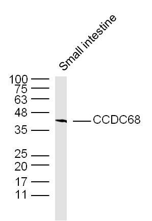
| Calculated MW | 39 KDa |
| Physical State | Liquid |
| Immunogen | KLH conjugated synthetic peptide derived from Human CCDC68/se57-1 |
| Epitope Specificity | 255-335/335 |
| Isotype | IgG |
| Purity | affinity purified by Protein A |
| Buffer | 0.01M TBS (pH7.4) with 1% BSA, 0.02% Proclin300 and 50% Glycerol. |
| Important Note | This product as supplied is intended for research use only, not for use in human, therapeutic or diagnostic applications. |
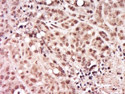
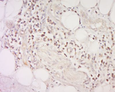
| Background Descriptions | CCDC68, also known as CTCL tumor antigen se57-1, is a 335 amino acid protein expressed in cutaneous T-cell lymphoma (CTCL), bone marrow, colon, small intestine, spleen, testis and trachea tissues. CTCL is a cancer of the immune system associated with mutation of the T cells. Malignant T cells form lesions on the surface of the skin. se57-1 contains 1 coiled coil domain and is encoded by the CCDC68 gene mapping to human chromosome 18. Chromosome 18 houses over 300 protein-coding genes and contains nearly 76 million bases. There are a variety of diseases associated with defects in chromosome 18-localized genes, some of which include Trisomy 18 (also known as Edwards syndrome), Niemann-Pick disease, hereditary hemorrhagic telangiectasia, erythropoietic protoporphyria and follicular lymphomas. |
| Gene ID | 80323 |
|---|---|
| Other Names | Coiled-coil domain-containing protein 68, Cutaneous T-cell lymphoma-associated antigen se57-1, CTCL-associated antigen se57-1, CCDC68 |
| Target/Specificity | Expressed in bone marrow, colon, small intestine, spleen, testis, trachea and cutaneous T-cell lymphoma (CTCL). |
| Dilution | WB=1:500-2000,IHC-P=1:100-500,IHC-F=1:100-500,IF=1:100-500,ELISA=1:5000-10000 |
| Storage | Store at -20 ℃ for one year. Avoid repeated freeze/thaw cycles. When reconstituted in sterile pH 7.4 0.01M PBS or diluent of antibody the antibody is stable for at least two weeks at 2-4 ℃. |
| Name | CCDC68 |
|---|---|
| Function | Centriolar protein required for centriole subdistal appendage assembly and microtubule anchoring in interphase cells (PubMed:28422092). Together with CCDC120, cooperate with subdistal appendage components ODF2, NIN and CEP170 for hierarchical subdistal appendage assembly (PubMed:28422092). |
| Cellular Location | Cytoplasm, cytoskeleton, microtubule organizing center, centrosome, centriole Note=Localizes to the subdistal appendages of centrioles (PubMed:28422092). |
| Tissue Location | Expressed in bone marrow, colon, small intestine, spleen, testis, trachea and cutaneous T-cell lymphoma (CTCL) |
Research Areas
Citations (0)

Thousands of laboratories across the world have published research that depended on the performance of antibodies from Abcepta to advance their research. Check out links to articles that cite our products in major peer-reviewed journals, organized by research category.
Submit your citation using an Abcepta antibody to
info@abcepta.com, and receive a free "I Love Antibodies" mug.
info@abcepta.com, and receive a free "I Love Antibodies" mug.

Application Protocols
Provided below are standard protocols that you may find useful for product applications.
Abcepta welcomes feedback from its customers.
If you have used an Abcepta product and would like to share how it has performed, please click on the "Submit Review" button and provide the requested information. Our staff will examine and post your review and contact you if needed.
If you have any additional inquiries please email technical services at tech@abcepta.com.
$ 385.00
Cat# AP58864
Ordering Information
United States
AlbaniaAustraliaAustriaBelgiumBosnia & HerzegovinaBrazilBulgariaCanadaCentral AmericaChinaCroatiaCyprusCzech RepublicDenmarkEstoniaFinlandFranceGermanyGreeceHong KongHungaryIcelandIndiaIndonesiaIrelandIsraelItalyJapanLatviaLithuaniaLuxembourgMacedoniaMalaysiaMaltaMexicoNetherlandsNew ZealandNorwayPakistanPolandPortugalRomaniaSerbiaSingaporeSlovakiaSloveniaSouth AfricaSouth KoreaSpainSwedenSwitzerlandTaiwanTurkeyUnited KingdomUnited StatesVietnamWorldwideOthers
USA Headquarters
(888) 735-7227 / (858) 622-0099 or (858) 875-1900
Shipping Information
Domestic orders (in stock items)
Shipped out the same day. Orders placed after 1 PM (PST) will ship out the next business day.
International orders
Contact your local distributors
