


 Foundational characteristics of cancer include proliferation, angiogenesis, migration, evasion of apoptosis, and cellular immortality. Find key markers for these cellular processes and antibodies to detect them.
 The SUMOplot™ Analysis Program predicts and scores sumoylation sites in your protein. SUMOylation is a post-translational modification involved in various cellular processes, such as nuclear-cytosolic transport, transcriptional regulation, apoptosis, protein stability, response to stress, and progression through the cell cycle.
 The Autophagy Receptor Motif Plotter predicts and scores autophagy receptor binding sites in your protein. Identifying proteins connected to this pathway is critical to understanding the role of autophagy in physiological as well as pathological processes such as development, differentiation, neurodegenerative diseases, stress, infection, and cancer.
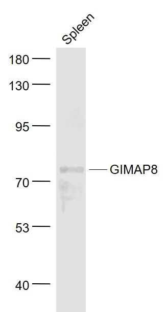
GIMAP8 Polyclonal Antibody
Purified Rabbit Polyclonal Antibody (Pab)
- SPECIFICATION
- CITATIONS
- PROTOCOLS
- BACKGROUND

Application
| WB, IHC-P, IHC-F, IF, E |
|---|---|
| Primary Accession | Q8ND71 |
| Reactivity | Rat |
| Host | Rabbit |
| Clonality | Polyclonal |
| Calculated MW | 75 KDa |
| Physical State | Liquid |
| Immunogen | KLH conjugated synthetic peptide derived from human GIMAP8 |
| Epitope Specificity | 551-565/665 |
| Isotype | IgG |
| Purity | affinity purified by Protein A |
| Buffer | 0.01M TBS (pH7.4) with 1% BSA, 0.02% Proclin300 and 50% Glycerol. |
| SUBCELLULAR LOCATION | Endoplasmic reticulum (By similarity). Golgi apparatus (By similarity). Mitochondrion (By similarity). |
| SIMILARITY | Belongs to the IAN GTP-binding protein family. |
| Important Note | This product as supplied is intended for research use only, not for use in human, therapeutic or diagnostic applications. |
| Background Descriptions | The GTPase of the immunity-associated protein (GIMAP) family of proteins include seven members that are expressed by genes residing on human chromosome 7. GIMAP proteins have been implicated in the regulation of lymphomyeloid cell survival. GIMAP8, also known as IAN9 (immune-associated nucleotide-binding protein 9) or IANT, is a 665 amino acid protein that localizes to Golgi apparatus, Endoplasmic reticulum and mitochondria. Suggested to have an anti-apoptotic effect on the immune system, GIMAP8 plays a role in infection response and is encoded by a gene that maps to human chromosome 7q36.1. Chromosome 7 houses over 1,000 genes, comprises nearly 5% of the human genome and has been linked to Osteogenesis imperfecta, Pendred syndrome, Lissencephaly, Citrullinemia and Shwachman-Diamond syndrome. |
| Gene ID | 155038 |
|---|---|
| Other Names | GTPase IMAP family member 8, Immune-associated nucleotide-binding protein 9, IAN-9, Protein IanT, GIMAP8, IAN9, IANT |
| Dilution | WB=1:500-2000,IHC-P=1:100-500,IHC-F=1:100-500,IF=1:100-500,ELISA=1:5000-10000 |
| Storage | Store at -20 ℃ for one year. Avoid repeated freeze/thaw cycles. When reconstituted in sterile pH 7.4 0.01M PBS or diluent of antibody the antibody is stable for at least two weeks at 2-4 ℃. |
| Name | GIMAP8 |
|---|---|
| Synonyms | IAN9, IANT |
| Function | Exerts an anti-apoptotic effect in the immune system and is involved in responses to infections. |
| Cellular Location | Endoplasmic reticulum {ECO:0000250|UniProtKB:Q75N62}. Golgi apparatus {ECO:0000250|UniProtKB:Q75N62}. Mitochondrion {ECO:0000250|UniProtKB:Q75N62}. Cytoplasm, cytosol |
| Tissue Location | Expressed in the spleen, intestine, liver, and colon, as well as in lung, placenta, kidney, muscle, and heart Extremely low expression, if any, in brain, in thymus, bone marrow, and blood leukocytes (PubMed:15474311). Detected in T-cells (PubMed:23454188). |
Citations (0)

Thousands of laboratories across the world have published research that depended on the performance of antibodies from Abcepta to advance their research. Check out links to articles that cite our products in major peer-reviewed journals, organized by research category.
Submit your citation using an Abcepta antibody to
info@abcepta.com, and receive a free "I Love Antibodies" mug.
info@abcepta.com, and receive a free "I Love Antibodies" mug.

Application Protocols
Provided below are standard protocols that you may find useful for product applications.
Abcepta welcomes feedback from its customers.
If you have used an Abcepta product and would like to share how it has performed, please click on the "Submit Review" button and provide the requested information. Our staff will examine and post your review and contact you if needed.
If you have any additional inquiries please email technical services at tech@abcepta.com.
$ 385.00
Cat# AP58950
Ordering Information
United States
AlbaniaAustraliaAustriaBelgiumBosnia & HerzegovinaBrazilBulgariaCanadaCentral AmericaChinaCroatiaCyprusCzech RepublicDenmarkEstoniaFinlandFranceGermanyGreeceHong KongHungaryIcelandIndiaIndonesiaIrelandIsraelItalyJapanLatviaLithuaniaLuxembourgMacedoniaMalaysiaMaltaMexicoNetherlandsNew ZealandNorwayPakistanPolandPortugalRomaniaSerbiaSingaporeSlovakiaSloveniaSouth AfricaSouth KoreaSpainSwedenSwitzerlandTaiwanTurkeyUnited KingdomUnited StatesVietnamWorldwideOthers
USA Headquarters
(888) 735-7227 / (858) 622-0099 or (858) 875-1900
Shipping Information
Domestic orders (in stock items)
Shipped out the same day. Orders placed after 1 PM (PST) will ship out the next business day.
International orders
Contact your local distributors
