


 Foundational characteristics of cancer include proliferation, angiogenesis, migration, evasion of apoptosis, and cellular immortality. Find key markers for these cellular processes and antibodies to detect them.
 The SUMOplot™ Analysis Program predicts and scores sumoylation sites in your protein. SUMOylation is a post-translational modification involved in various cellular processes, such as nuclear-cytosolic transport, transcriptional regulation, apoptosis, protein stability, response to stress, and progression through the cell cycle.
 The Autophagy Receptor Motif Plotter predicts and scores autophagy receptor binding sites in your protein. Identifying proteins connected to this pathway is critical to understanding the role of autophagy in physiological as well as pathological processes such as development, differentiation, neurodegenerative diseases, stress, infection, and cancer.
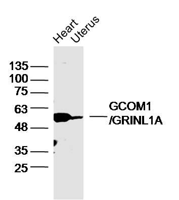
GCOM1/GRINL1A Polyclonal Antibody
Purified Rabbit Polyclonal Antibody (Pab)
- SPECIFICATION
- CITATIONS
- PROTOCOLS
- BACKGROUND

Application
| WB, IHC-P, IHC-F, IF, E |
|---|---|
| Primary Accession | P0CAP2 |
| Reactivity | Rat, Bovine |
| Host | Rabbit |
| Clonality | Polyclonal |
| Calculated MW | 62 KDa |
| Physical State | Liquid |
| Immunogen | KLH conjugated synthetic peptide derived from human GCOM1 |
| Epitope Specificity | 51-150/550 |
| Isotype | IgG |
| Purity | affinity purified by Protein A |
| Buffer | 0.01M TBS (pH7.4) with 1% BSA, 0.02% Proclin300 and 50% Glycerol. |
| Important Note | This product as supplied is intended for research use only, not for use in human, therapeutic or diagnostic applications. |
| Background Descriptions | Glutamate receptors mediate most excitatory neurotransmission in the brain and play an important role in neural plasticity, neural development and neurodegeneration. Ionotropic glutamate receptors are categorized into NMDA receptors and kainate/AMPA receptors, both of which contain glutamate-gated, cation-specific ion channels. Synaptic and extrasynaptic NMDA receptors have been shown to have opposite effects on neuronal survival, CREB function and gene regulation. Gcom1 (GRINL1A complex locus protein 1), also known as GUP (GRINL1A upstream protein) and Gcom (GRINL1A combined protein), is a 466 amino acid protein that is a component of the GRINL1A complex transcription unit, which is thought to be involved in the modulation of glutamatergic neurotransmission through interaction with the NR1 subunit of the NMDA receptor. Gcom1 is expressed in small intestine, lung, liver, heart, skeletal muscle, testis and prostate and also colocalizes with NR1 in cortical and hippocampal neurons. There are eleven isoforms of Gcom1 that are produced as a result of alternative splicing events. |
| Gene ID | 81488 |
|---|---|
| Other Names | DNA-directed RNA polymerase II subunit GRINL1A, DNA-directed RNA polymerase II subunit M, Glutamate receptor-like protein 1A, POLR2M, GRINL1A |
| Dilution | WB=1:500-2000,IHC-P=1:100-500,IHC-F=1:100-500,IF=1:50-200,ELISA=1:5000-10000 |
| Storage | Store at -20 ℃ for one year. Avoid repeated freeze/thaw cycles. When reconstituted in sterile pH 7.4 0.01M PBS or diluent of antibody the antibody is stable for at least two weeks at 2-4 ℃. |
| Name | POLR2M |
|---|---|
| Synonyms | GRINL1A |
| Function | [Isoform 1]: Appears to be a stable component of the Pol II(G) complex form of RNA polymerase II (Pol II). Pol II synthesizes mRNA precursors and many functional non-coding RNAs and is the central component of the basal RNA polymerase II transcription machinery. May play a role in the Mediator complex-dependent regulation of transcription activation. Acts as a negative regulator of transcriptional activation; this repression is relieved by the Mediator complex, which restores Pol II(G) activator-dependent transcription to a level equivalent to that of Pol II. |
| Cellular Location | [Isoform 1]: Nucleus. |
| Tissue Location | Detected in adult an fetal brain. Detected in heart, kidney, skeletal muscle, small intestine, lung, prostate and testis. |
Research Areas
Citations (0)

Thousands of laboratories across the world have published research that depended on the performance of antibodies from Abcepta to advance their research. Check out links to articles that cite our products in major peer-reviewed journals, organized by research category.
Submit your citation using an Abcepta antibody to
info@abcepta.com, and receive a free "I Love Antibodies" mug.
info@abcepta.com, and receive a free "I Love Antibodies" mug.

Application Protocols
Provided below are standard protocols that you may find useful for product applications.
Abcepta welcomes feedback from its customers.
If you have used an Abcepta product and would like to share how it has performed, please click on the "Submit Review" button and provide the requested information. Our staff will examine and post your review and contact you if needed.
If you have any additional inquiries please email technical services at tech@abcepta.com.
$ 385.00
Cat# AP58991
Ordering Information
United States
AlbaniaAustraliaAustriaBelgiumBosnia & HerzegovinaBrazilBulgariaCanadaCentral AmericaChinaCroatiaCyprusCzech RepublicDenmarkEstoniaFinlandFranceGermanyGreeceHong KongHungaryIcelandIndiaIndonesiaIrelandIsraelItalyJapanLatviaLithuaniaLuxembourgMacedoniaMalaysiaMaltaMexicoNetherlandsNew ZealandNorwayPakistanPolandPortugalRomaniaSerbiaSingaporeSlovakiaSloveniaSouth AfricaSouth KoreaSpainSwedenSwitzerlandTaiwanTurkeyUnited KingdomUnited StatesVietnamWorldwideOthers
USA Headquarters
(888) 735-7227 / (858) 622-0099 or (858) 875-1900
Other Products
Shipping Information
Domestic orders (in stock items)
Shipped out the same day. Orders placed after 1 PM (PST) will ship out the next business day.
International orders
Contact your local distributors
