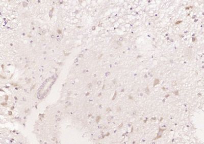
LRSAM1 Polyclonal Antibody
Purified Rabbit Polyclonal Antibody (Pab)
- SPECIFICATION
- CITATIONS
- PROTOCOLS
- BACKGROUND

Application
| WB, IHC-P, IHC-F, IF, E |
|---|---|
| Primary Accession | Q6UWE0 |
| Reactivity | Rat, Bovine |
| Host | Rabbit |
| Clonality | Polyclonal |
| Calculated MW | 84 KDa |
| Physical State | Liquid |
| Immunogen | KLH conjugated synthetic peptide derived from human LRSAM1 |
| Epitope Specificity | 201-300/723 |
| Isotype | IgG |
| Purity | affinity purified by Protein A |
| Buffer | 0.01M TBS (pH7.4) with 1% BSA, 0.02% Proclin300 and 50% Glycerol. |
| SUBCELLULAR LOCATION | Cytoplasm. Note=Displays a punctuate distribution and localizes to a submembranal ring. |
| SIMILARITY | Contains 6 LRR (leucine-rich) repeats. Contains 1 RING-type zinc finger. Contains 1 SAM (sterile alpha motif) domain. |
| SUBUNIT | Interacts with TSG101. |
| DISEASE | Defects in LRSAM1 are a cause of Charcot-Marie-Tooth disease type 2P (CMT2P) [MIM:614436]. CMT2P is an axonal form of Charcot-Marie-Tooth disease, a disorder of the peripheral nervous system, characterized by progressive weakness and atrophy, initially of the peroneal muscles and later of the distal muscles of the arms. Charcot-Marie-Tooth disease is classified in two main groups on the basis of electrophysiologic properties and histopathology: primary peripheral demyelinating neuropathies (designated CMT1 when they are dominantly inherited) and primary peripheral axonal neuropathies (CMT2). Neuropathies of the CMT2 group are characterized by signs of axonal degeneration in the absence of obvious myelin alterations, normal or slightly reduced nerve conduction velocities, and progressive distal muscle weakness and atrophy. Nerve conduction velocities are normal or slightly reduced. |
| Important Note | This product as supplied is intended for research use only, not for use in human, therapeutic or diagnostic applications. |
| Background Descriptions | LRSAM1 is an E3 ubiquitin-protein ligase that mediates monoubiquitination of TSG101 at multiple sites, leading to inactivation of the ability of TSG101 to sort endocytic (EGF receptors) and exocytic (HIV-1 viral proteins) cargos. It selectively regulates cell adhesion molecules and plays a role in receptor endocytosis and viral budding. LRSAM1 contains a RING-type zinc finger, 5 leucine-rich repeats and 1 SAM (sterile alpha motif) domain. The coiled coil domains interact with the SB domain of TSG101. The PTAP motifs mediate the binding to UEV domains. There are 3 isoforms produced by alternative splicing. |
| Gene ID | 90678 |
|---|---|
| Other Names | E3 ubiquitin-protein ligase LRSAM1, 2.3.2.27, Leucine-rich repeat and sterile alpha motif-containing protein 1, RING-type E3 ubiquitin transferase LRSAM1, Tsg101-associated ligase, hTAL, LRSAM1 {ECO:0000303|PubMed:20865121} |
| Target/Specificity | Highly expressed in adult spinal cord motoneurons as well as in fetal spinal cord and muscle tissue. |
| Dilution | WB=1:500-2000,IHC-P=1:100-500,IHC-F=1:100-500,IF=1:50-200,ELISA=1:5000-10000 |
| Storage | Store at -20 ℃ for one year. Avoid repeated freeze/thaw cycles. When reconstituted in sterile pH 7.4 0.01M PBS or diluent of antibody the antibody is stable for at least two weeks at 2-4 ℃. |
| Name | LRSAM1 {ECO:0000303|PubMed:20865121} |
|---|---|
| Function | E3 ubiquitin-protein ligase that mediates monoubiquitination of TSG101 at multiple sites, leading to inactivate the ability of TSG101 to sort endocytic (EGF receptors) and exocytic (HIV-1 viral proteins) cargos (PubMed:15256501). Bacterial recognition protein that defends the cytoplasm from invasive pathogens (PubMed:23245322). Localizes to several intracellular bacterial pathogens and generates the bacteria-associated ubiquitin signal leading to autophagy-mediated intracellular bacteria degradation (xenophagy) (PubMed:23245322, PubMed:25484098). |
| Cellular Location | Cytoplasm. Note=Displays a punctuate distribution and localizes to a submembranal ring (PubMed:15256501). Localizes to intracellular bacterial pathogens (PubMed:23245322) |
| Tissue Location | Highly expressed in adult spinal cord motoneurons as well as in fetal spinal cord and muscle tissue |

Thousands of laboratories across the world have published research that depended on the performance of antibodies from Abcepta to advance their research. Check out links to articles that cite our products in major peer-reviewed journals, organized by research category.
info@abcepta.com, and receive a free "I Love Antibodies" mug.
Provided below are standard protocols that you may find useful for product applications.
If you have used an Abcepta product and would like to share how it has performed, please click on the "Submit Review" button and provide the requested information. Our staff will examine and post your review and contact you if needed.
If you have any additional inquiries please email technical services at tech@abcepta.com.


 Foundational characteristics of cancer include proliferation, angiogenesis, migration, evasion of apoptosis, and cellular immortality. Find key markers for these cellular processes and antibodies to detect them.
 The SUMOplot™ Analysis Program predicts and scores sumoylation sites in your protein. SUMOylation is a post-translational modification involved in various cellular processes, such as nuclear-cytosolic transport, transcriptional regulation, apoptosis, protein stability, response to stress, and progression through the cell cycle.
 The Autophagy Receptor Motif Plotter predicts and scores autophagy receptor binding sites in your protein. Identifying proteins connected to this pathway is critical to understanding the role of autophagy in physiological as well as pathological processes such as development, differentiation, neurodegenerative diseases, stress, infection, and cancer.


