


 Foundational characteristics of cancer include proliferation, angiogenesis, migration, evasion of apoptosis, and cellular immortality. Find key markers for these cellular processes and antibodies to detect them.
 The SUMOplot™ Analysis Program predicts and scores sumoylation sites in your protein. SUMOylation is a post-translational modification involved in various cellular processes, such as nuclear-cytosolic transport, transcriptional regulation, apoptosis, protein stability, response to stress, and progression through the cell cycle.
 The Autophagy Receptor Motif Plotter predicts and scores autophagy receptor binding sites in your protein. Identifying proteins connected to this pathway is critical to understanding the role of autophagy in physiological as well as pathological processes such as development, differentiation, neurodegenerative diseases, stress, infection, and cancer.
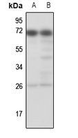
Anti-TESK1 Antibody
Rabbit polyclonal antibody to TESK1
- SPECIFICATION
- CITATIONS
- PROTOCOLS
- BACKGROUND

Application
| WB, IP |
|---|---|
| Primary Accession | Q15569 |
| Reactivity | Human, Rat, Monkey |
| Host | Rabbit |
| Clonality | Polyclonal |
| Calculated MW | 67684 Da |
| Gene ID | 7016 |
|---|---|
| Other Names | Dual specificity testis-specific protein kinase 1; Testicular protein kinase 1 |
| Target/Specificity | KLH-conjugated synthetic peptide encompassing a sequence within the center region of human TESK1. The exact sequence is proprietary. |
| Dilution | WB~~WB (1/500 - 1/1000), IP (1/10 - 1/100) IP~~N/A |
| Format | Liquid in 0.42% Potassium phosphate, 0.87% Sodium chloride, pH 7.3, 30% glycerol, and 0.09% (W/V) sodium azide. |
| Storage | Store at -20 °C.Stable for 12 months from date of receipt |
| Name | TESK1 |
|---|---|
| Function | Dual specificity protein kinase activity catalyzing autophosphorylation and phosphorylation of exogenous substrates on both serine/threonine and tyrosine residues (By similarity). Regulates the cellular cytoskeleton by enhancing actin stress fiber formation via phosphorylation of cofilin and by preventing microtubule breakdown via inhibition of TAOK1/MARKK kinase activity (By similarity). Inhibits podocyte motility via regulation of actin cytoskeletal dynamics and phosphorylation of CFL1 (By similarity). Positively regulates integrin- mediated cell spreading, via phosphorylation of cofilin (PubMed:15584898). Suppresses ciliogenesis via multiple pathways; phosphorylation of CFL1, suppression of ciliary vesicle directional trafficking to the ciliary base, and by facilitating YAP1 nuclear localization where it acts as a transcriptional corepressor of the TEAD4 target genes AURKA and PLK1 (PubMed:25849865). Probably plays a central role at and after the meiotic phase of spermatogenesis (By similarity). |
| Cellular Location | Cytoplasm. Cytoplasm, perinuclear region {ECO:0000250|UniProtKB:Q63572} Cytoplasm, cytoskeleton, microtubule organizing center, centrosome. Cell projection, lamellipodium {ECO:0000250|UniProtKB:Q63572}. Note=Colocalizes with SPRY4 in vesicular spots in the cytoplasm (PubMed:15584898). Localized to F- actin-rich lamellipodia at the cell periphery following fibronectin- mediated cell adhesion of Schwann cells (By similarity) {ECO:0000250|UniProtKB:Q63572, ECO:0000269|PubMed:15584898} |
| Tissue Location | Expressed in podocytes and renal tubular cells in the kidney (at protein level). |

Thousands of laboratories across the world have published research that depended on the performance of antibodies from Abcepta to advance their research. Check out links to articles that cite our products in major peer-reviewed journals, organized by research category.
info@abcepta.com, and receive a free "I Love Antibodies" mug.

Provided below are standard protocols that you may find useful for product applications.
Background
KLH-conjugated synthetic peptide encompassing a sequence within the center region of human TESK1. The exact sequence is proprietary.
If you have used an Abcepta product and would like to share how it has performed, please click on the "Submit Review" button and provide the requested information. Our staff will examine and post your review and contact you if needed.
If you have any additional inquiries please email technical services at tech@abcepta.com.
Ordering Information
Other Products
Shipping Information
