


 Foundational characteristics of cancer include proliferation, angiogenesis, migration, evasion of apoptosis, and cellular immortality. Find key markers for these cellular processes and antibodies to detect them.
 The SUMOplot™ Analysis Program predicts and scores sumoylation sites in your protein. SUMOylation is a post-translational modification involved in various cellular processes, such as nuclear-cytosolic transport, transcriptional regulation, apoptosis, protein stability, response to stress, and progression through the cell cycle.
 The Autophagy Receptor Motif Plotter predicts and scores autophagy receptor binding sites in your protein. Identifying proteins connected to this pathway is critical to understanding the role of autophagy in physiological as well as pathological processes such as development, differentiation, neurodegenerative diseases, stress, infection, and cancer.
Anti-TRPV3 Antibody
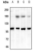
Rabbit polyclonal antibody to TRPV3
- SPECIFICATION
- CITATIONS
- PROTOCOLS
- BACKGROUND

Application
| WB |
|---|---|
| Primary Accession | Q8NET8 |
| Reactivity | Human, Mouse, Rat |
| Host | Rabbit |
| Clonality | Polyclonal |
| Calculated MW | 90636 Da |
| Gene ID | 162514 |
|---|---|
| Other Names | Transient receptor potential cation channel subfamily V member 3; TrpV3; Vanilloid receptor-like 3; VRL-3 |
| Target/Specificity | KLH-conjugated synthetic peptide encompassing a sequence within the center region of human TRPV3. The exact sequence is proprietary. |
| Dilution | WB~~WB (1/500 - 1/1000) |
| Format | Liquid in 0.42% Potassium phosphate, 0.87% Sodium chloride, pH 7.3, 30% glycerol, and 0.09% (W/V) sodium azide. |
| Storage | Store at -20 °C.Stable for 12 months from date of receipt |
| Name | TRPV3 |
|---|---|
| Function | Non-selective calcium permeant cation channel (PubMed:12077604, PubMed:12077606, PubMed:26818531, PubMed:37648856, PubMed:38691614). It is activated by innocuous (warm) temperatures and shows an increased response at noxious temperatures greater than 39 degrees Celsius (PubMed:12077604, PubMed:12077606). Activation exhibits an outward rectification (PubMed:12077604). The channel pore can dilate to provide permeability to larger cations (PubMed:37648856). May associate with TRPV1 and may modulate its activity (PubMed:12077606). Is a negative regulator of hair growth and cycling: TRPV3-coupled signaling suppresses keratinocyte proliferation in hair follicles and induces apoptosis and premature hair follicle regression (catagen) (PubMed:21593771). |
| Cellular Location | Cell membrane; Multi-pass membrane protein. Cytoplasm Lysosome. Note=Targeted to lysosome for degradation in a SNX11-dependent manner. |
| Tissue Location | Abundantly expressed in CNS. Widely expressed at low levels. Detected in dorsal root ganglion (at protein level) Expressed in the keratinocyte layers of the outer root sheath and, to lesser extent, to the matrix of the hair follicles (at protein level) |

Thousands of laboratories across the world have published research that depended on the performance of antibodies from Abcepta to advance their research. Check out links to articles that cite our products in major peer-reviewed journals, organized by research category.
info@abcepta.com, and receive a free "I Love Antibodies" mug.

Provided below are standard protocols that you may find useful for product applications.
Background
KLH-conjugated synthetic peptide encompassing a sequence within the center region of human TRPV3. The exact sequence is proprietary.
If you have used an Abcepta product and would like to share how it has performed, please click on the "Submit Review" button and provide the requested information. Our staff will examine and post your review and contact you if needed.
If you have any additional inquiries please email technical services at tech@abcepta.com.
Ordering Information
Other Products
Shipping Information
